. 2021 Jul;38(4):794-799.
doi: 10.1111/pde.14624. Epub 2021 Jun 8.

Dermatologic findings in individuals with genetically confirmed Proteus syndrome

Affiliations

Dermatologic findings in individuals with genetically confirmed Proteus syndrome

Deeti J Pithadia et al. Pediatr Dermatol. 2021 Jul.

Abstract

Background/objective: Proteus syndrome, caused by a mosaic activating AKT1 variant, typically presents in toddlers with progressive, asymmetric overgrowth of the skin and bones. We aimed to define the spectrum of dermatologic disease in individuals with genetically confirmed Proteus syndrome.

Methods: We conducted a retrospective review of records from dermatologic examinations of individuals evaluated at the NIH with a molecular diagnosis of Proteus syndrome. The types, prevalence, and localization of dermatologic findings were assessed.

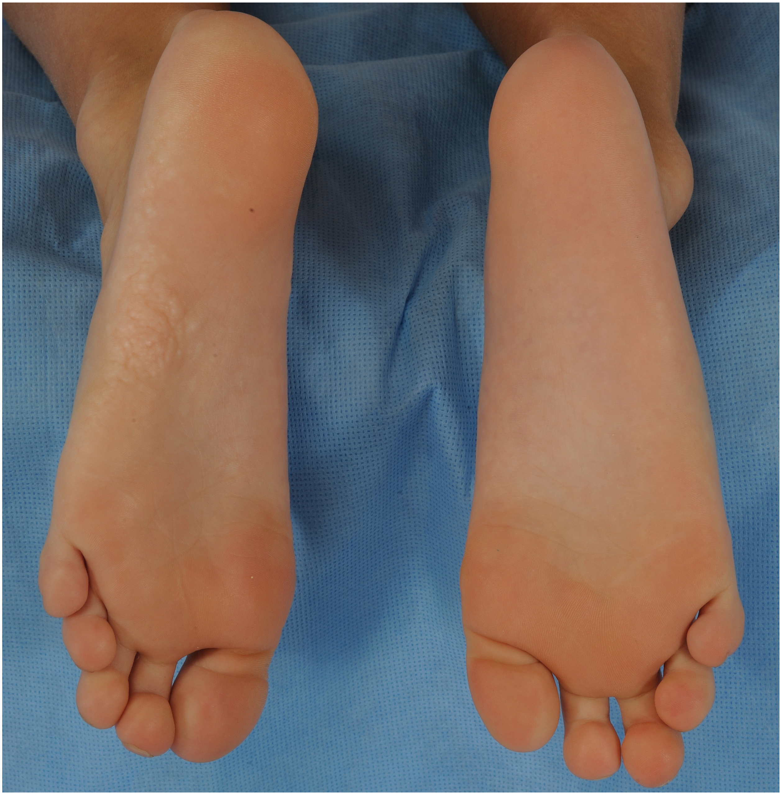
Results: Fifty-one individuals (29 males, 22 females, mean age: 9 years) with clinical features of Proteus syndrome had the mosaic c.49G>A, p.Glu17Lys AKT1 variant. Fifty (98%) had at least one cutaneous feature constituting current clinical diagnostic criteria, including vascular malformations in 42 (82%), epidermal nevus in 41 (80%), volar cerebriform connective tissue nevi in 34 (67%), and adipose dysregulation in 30 (59%). Forty-nine (96%) had at least one dermatologic finding not included within the diagnostic criteria, including confluent volar skin-colored to hypopigmented papules or nodules (n = 33, 65%), papules or nodules on the digits or face (n = 27, 53%), and nonlinear epidermal nevi (n = 15, 29%). Other frequently observed features include nail changes (n = 28, 55%), hyperpigmented macules (n = 27, 53%), patchy dermal hypoplasia (n = 18, 35%), gingival/oral mucosal overgrowth (n = 17, 33%), hypopigmented macules (n = 16, 31%), dental enamel changes (n = 9, 18%), acrochordons (n = 6, 12%), and lingual overgrowth (n = 4, 8%).

Conclusions: The range of mucocutaneous features occurring in Proteus syndrome is broader than previously considered. These observations may assist in earlier diagnosis and management and provide novel insights regarding the pathogenesis of the condition.

Keywords: Genetic disease/mechanisms; genodermatoses; neoplasms-benign; skin signs of systemic disease; vascular malformations.


Figures

Figure 1:
Figure 1:
Development of the cerebriform connective tissue nevus in Proteus syndrome. A. A 5-year-old boy has a plaque on his left sole consisting of confluent, slightly hypopigmented papules. B. At the age of 13 years, the plaque on the left foot has expanded and developed into a characteristic cerebriform connective tissue nevus, while the sole of the right foot has developed a plaque with a few areas showing the beginnings of cerebriform morphology.
Figure 1:
Figure 1:
Development of the cerebriform connective tissue nevus in Proteus syndrome. A. A 5-year-old boy has a plaque on his left sole consisting of confluent, slightly hypopigmented papules. B. At the age of 13 years, the plaque on the left foot has expanded and developed into a characteristic cerebriform connective tissue nevus, while the sole of the right foot has developed a plaque with a few areas showing the beginnings of cerebriform morphology.
Figure 2:
Figure 2:
Papular and nodular connective tissue nevi in Proteus syndrome. A. The only lesions of a connective tissue nevus in this 14-year-old girl were a papule on the left fourth toe and a nodule on the left fifth toe. Biopsy of the nodule confirmed connective tissue nevus and the presence of the AKT1 variant. B. Firm, skin-colored papules and nodules on the lower eyelid, perinasal region, and the lower vermilion border in an 8-year-old boy.
Figure 2:
Figure 2:
Papular and nodular connective tissue nevi in Proteus syndrome. A. The only lesions of a connective tissue nevus in this 14-year-old girl were a papule on the left fourth toe and a nodule on the left fifth toe. Biopsy of the nodule confirmed connective tissue nevus and the presence of the AKT1 variant. B. Firm, skin-colored papules and nodules on the lower eyelid, perinasal region, and the lower vermilion border in an 8-year-old boy.
Figure 3:
Figure 3:
Variations in appearance of epidermal nevus in Proteus syndrome. A. Verrucous nonlinear epidermal nevus on the back of the right hand, with a surgical scar. B. Epidermal nevus on the lateral trunk with a thick, papillomatous or acrochordon-like appearance and terminal hair growth in a 17-year-old boy.
Figure 3:
Figure 3:
Variations in appearance of epidermal nevus in Proteus syndrome. A. Verrucous nonlinear epidermal nevus on the back of the right hand, with a surgical scar. B. Epidermal nevus on the lateral trunk with a thick, papillomatous or acrochordon-like appearance and terminal hair growth in a 17-year-old boy.
Figure 4:
Figure 4:
Hyper- and hypopimented macules in a 12-year-old boy with Proteus syndrome. A. On the right lower back, there is a light brown macule with irregular borders. B. On the right upper back of the same child, there is a faint hypopigmented macule with a mottled pattern.
Figure 4:
Figure 4:
Hyper- and hypopimented macules in a 12-year-old boy with Proteus syndrome. A. On the right lower back, there is a light brown macule with irregular borders. B. On the right upper back of the same child, there is a faint hypopigmented macule with a mottled pattern.
Figure 5:
Figure 5:
Asymmetric gingival overgrowth, aberrancies in tooth shape, and abnormal dental enamel layer with exposed yellowish dentin in a 5-year-old girl with Proteus syndrome.

References

    1. Biesecker LG, Sapp JC. Proteus Syndrome. 2012August9 [Updated 2019 Jan 10]. In: Adam MP, Ardinger HH, Pagon RA, et al., editors. GeneReviews® [Internet]. Seattle (WA): University of Washington, Seattle; 1993–2021. Available from: https://www.ncbi.nlm.nih.gov/books/NBK99495/. - PubMed
    1. Nguyen D, Turner JT, Olsen C, Biesecker LG, Darling TN. Cutaneous manifestations of proteus syndrome: correlations with general clinical severity. Arch Dermatol. 2004;140(8):947–953. - PubMed
    1. Pithadia DJ RJ, Sapp JC, Biesecker LG, Darling TN. Hypertrichotic patches as a mosaic manifestation of Proteus syndrome. J Am Acad Dermatol. 2021;84(2):415–424. - PMC - PubMed
    1. Lindhurst MJ, Sapp JC, Teer JK, et al.A mosaic activating mutation in AKT1 associated with the Proteus syndrome. N Engl J Med. 2011;365(7):611–619. - PMC - PubMed
    1. Keppler-Noreuil KM, Sapp JC, Lindhurst MJ, et al.Pharmacodynamic Study of Miransertib in Individuals with Proteus Syndrome. Am J Hum Genet. 2019;104(3):484–491. - PMC - PubMed