Review
. 2021 Jun;39 Suppl 1(Suppl 1):15-23.
doi: 10.1002/hon.2857.

Langerhans cell histiocytosis: Version 2021

Affiliations
Review

Langerhans cell histiocytosis: Version 2021

Nitya Gulati et al. Hematol Oncol. 2021 Jun.

Abstract

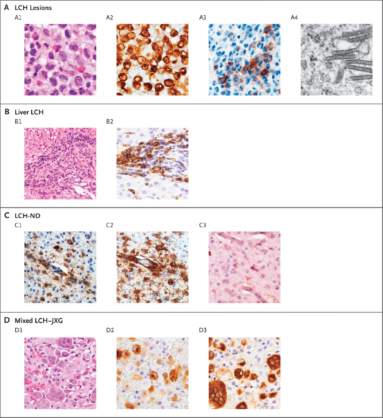
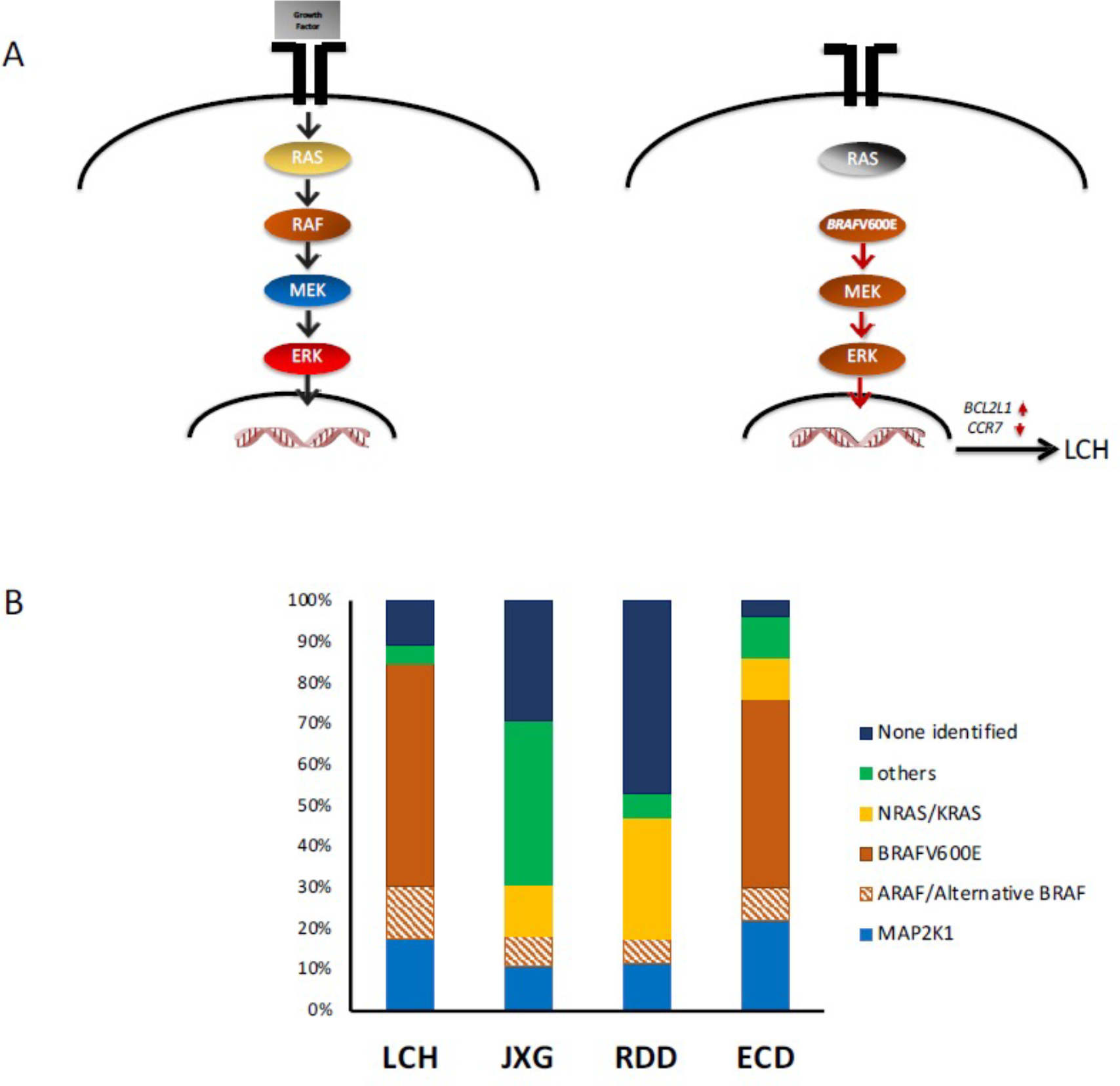
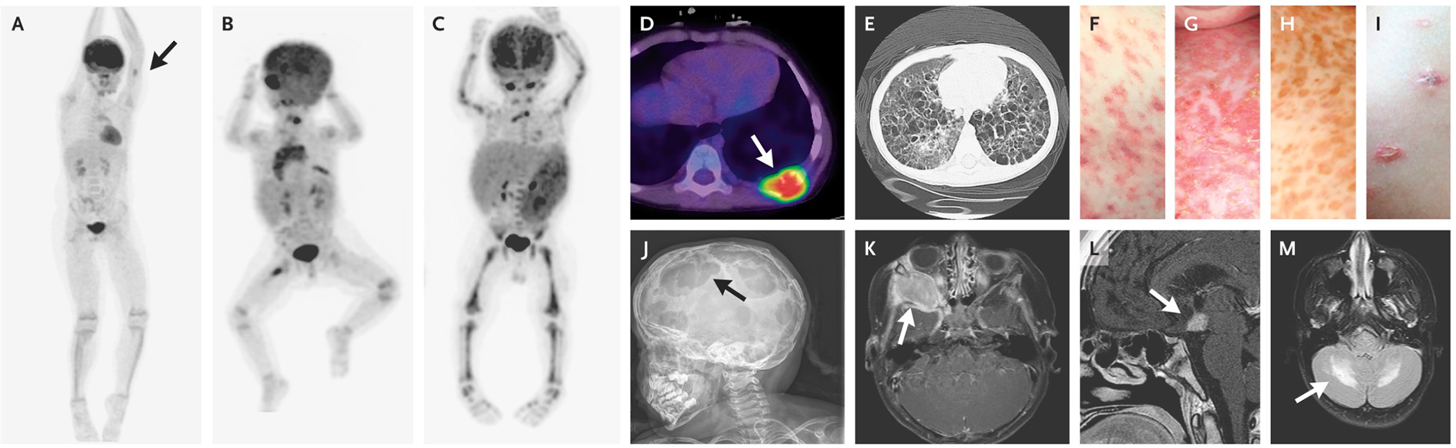
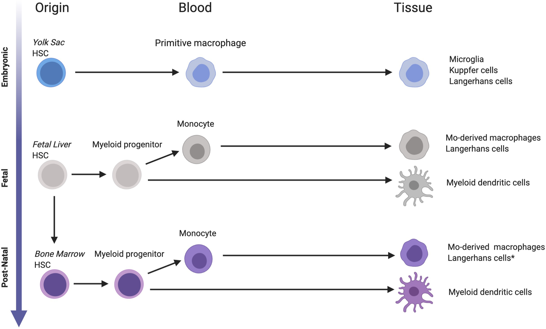
Children with Langerhnans cell histiocytosis (LCH) develop granulomatous lesions with characteristic clonal CD207+ dendritic cells that can arise as single lesions or life-threatening disseminated disease. Despite the wide range of clinical presentations, LCH lesions are histologically indistinguishable based on severity of disease, and uncertain classification as an immune versus neoplastic disorder has historically challenged the development of optimal clinical strategies for patients with LCH. Recently, activating somatic mutations in MAPK pathway genes, most notably BRAFV600E, have been discovered in almost all cases of LCH. Further, the stage of myeloid differentiation in which the mutation arises defines the extent of disease and risk of developing LCH-associated neurodegeneration. MAPK activation in LCH precursor cells drives myeloid differentiation, inhibits migration, and inhibits apoptosis, resulting in accumulation of resilient pathologic dendritic cells that recruit and activate T cells. Recurrent somatic mutations in MAPK pathway genes have also been identified in related histiocytic disorders: juvenile xanthogranuloma, Erdheim-Chester disease, and Rosai-Dorfman disease. New insights into pathogenesis support reclassification of these conditions as a myeloid neoplastic disorders. Continued research will uncover opportunities to identify novel targets and inform personalized therapeutic strategies based on cell of origin, somatic mutation, inherited risk factors, and residual disease.

Keywords: Erdheim-Chester disease; Langerhans cell histiocytosis; Rosai-Dorfman disease; histiocytic disorder; juvenile xanthogranuloma.


Conflict of interest statement

Conflict of Interest: The authors have no conflicts to report with respect to this manuscript.

Figures

Figure 1.
Figure 1.. Histologic Features of LCH.
Panel A shows typical LCH lesions with large cells, pale cytoplasm, and reniform nuclei on hematoxylin and eosin staining (A1); CD207-positive immunostaining (A2); VE1-positive immunostaining for BRAF V600E protein (A3); and Birbeck granules visualized with electron microscopy (A4). Panel B shows liver involvement, which is frequently characterized by periportal infiltration by histiocytes (B1) and variable CD207-positive staining (B2). Panel C shows biopsy specimens from a patient with severe LCH-associated neurodegeneration (LCH-ND), characterized by perivascular VE1-positive staining (C1), CD163-positive staining (C2), and a P2RY12 infiltrate with occasional P2RY12-positive, tissue-resident microglia (C3). Panel D shows histiocytic lesions that are characteristic of both LCH and juvenile xanthogranuloma (JXG), with heterogeneous histologic features on hematoxylin and eosin staining (D1), including distinct cell populations that are CD207-positive (D2) and CD68-positive (D3). (From NEJM, Langerhans Cell Histiocytosis, 379:856–868; Copyright ©2018 Massachusetts Medical Society. Reprinted with permission.)
Figure 2.
Figure 2.. MAPK Pathway Mutations in Histiocytic Disorders.
A. (left) Schematic of MAPK pathway. Under physiologic conditions, the growth factor (gray box) engages the tyrosine kinase receptor that transduces the signal to the nucleus. (right) Activating mutations (such as BRAF-V600E) drive constitutive ERK activation. In the case of LCH, this drives the expression of anti-apoptosis BCL2L1 (BCL-xL) and inhibits CCR7. B. Stacked bar graphs represent percentages of MAPK pathway mutations in each histiocytic histologic subtype.
Figure 3.
Figure 3.. Ontogeny of tissue “histiocytes”.
Tissue macrophages and dendritic cells seed tissues at various stages of development. Microglia, Küpffer cells and Langerhans cells arise from yolk sac-derived progenitors, followed by fetal liver, then adult hematopoiesis. *Langerhans cells may be generated from adult HSC monocyte-derived LCs following inflammation or tissue damage.
Figure 4.
Figure 4.. Clinical Presentations of LCH.
Positron-emission tomographic (PET) images show a single bone lesion involving the humerus (Panel A, arrow); low-risk lesions involving the orbit, lymph nodes, bone (multifocal lesion), and thymus (Panel B); and high-risk lesions involving the liver, spleen, and bone marrow (Panel C). Other classic presentations include a lytic bone lesion (Panel D, arrow), cystic lung lesions (Panel E), and various skin lesions (Panels F through I). Examples of LCH lesions involving the skull and brain include multifocal skull lesions (Panel J, arrow), an orbital lesion (Panel K, arrow), a pituitary lesion (Panel L, arrow), and LCH-associated neurodegeneration (Panel M, arrow). (From NEJM, Langerhans Cell Histiocytosis, 379:856–868; Copyright ©2018 Massachusetts Medical Society. Reprinted with permission.)

References

    1. Favara BE, Feller AC, Pauli M et al. Contemporary classification of histiocytic disorders. The WHO Committee On Histiocytic/Reticulum Cell Proliferations. Reclassification Working Group of the Histiocyte Society. Med Pediatr Oncol 1997;29:157–166. - PubMed
    1. Emile JF, Abla O, Fraitag S et al. Revised classification of histiocytoses and neoplasms of the macrophage-dendritic cell lineages. Blood 2016. - PMC - PubMed
    1. Allen CE, Merad M, McClain KL. Langerhans-Cell Histiocytosis. N Engl J Med 2018;379:856–868. - PMC - PubMed
    1. Badalian-Very G, Vergilio JA, Degar BA et al. Recurrent BRAF mutations in Langerhans cell histiocytosis. Blood 2010;116:1919–1923. - PMC - PubMed
    1. Durham BH, Lopez RE, Picarsic J et al. Activating mutations in CSF1R and additional receptor tyrosine kinases in histiocytic neoplasms. Nat Med 2019;25:1839–1842. - PMC - PubMed

MeSH terms

Substances