. 2021 Jun 1;148(11):dev199385.
doi: 10.1242/dev.199385. Epub 2021 Jun 9.

A patient-based medaka alg2 mutant as a model for hypo-N-glycosylation

Affiliations

A patient-based medaka alg2 mutant as a model for hypo-N-glycosylation

Sevinç Gücüm et al. Development. .

Abstract

Defects in the evolutionarily conserved protein-glycosylation machinery during embryonic development are often fatal. Consequently, congenital disorders of glycosylation (CDG) in human are rare. We modelled a putative hypomorphic mutation described in an alpha-1,3/1,6-mannosyltransferase (ALG2) index patient (ALG2-CDG) to address the developmental consequences in the teleost medaka (Oryzias latipes). We observed specific, multisystemic, late-onset phenotypes, closely resembling the patient's syndrome, prominently in the facial skeleton and in neuronal tissue. Molecularly, we detected reduced levels of N-glycans in medaka and in the patient's fibroblasts. This hypo-N-glycosylation prominently affected protein abundance. Proteins of the basic glycosylation and glycoprotein-processing machinery were over-represented in a compensatory response, highlighting the regulatory topology of the network. Proteins of the retinal phototransduction machinery, conversely, were massively under-represented in the alg2 model. These deficiencies relate to a specific failure to maintain rod photoreceptors, resulting in retinitis pigmentosa characterized by the progressive loss of these photoreceptors. Our work has explored only the tip of the iceberg of N-glycosylation-sensitive proteins, the function of which specifically impacts on cells, tissues and organs. Taking advantage of the well-described human mutation has allowed the complex interplay of N-glycosylated proteins and their contribution to development and disease to be addressed.

Keywords: CDG; Disease model; Glycosylation; Human hypomorphic mutations; Medaka; Retinitis pigmentosa.


Conflict of interest statement

Competing interests The authors declare no competing or financial interests.

Figures

Fig. 1.
Fig. 1.
Homozygous medaka alg2p.G336*/p.G336* mutants recapitulate ALG2-CDG multisystemic phenotype. (A) Schematic of human ALG2 and the medaka orthologous alg2 locus. The site of the ALG2 index patient mutation (c.1040delG) and targeted CRISPR genome editing (scissors) in medaka are indicated (dashed line). Single-stranded oligodeoxynucleotide (ssODN) donor for introduction of premature STOP (asterisk) is given. Red box, coding exon; white box, untranslated region. (A′) Predicted protein variants at the given position of human ALG2 index patient and corresponding sites of the stable mutant fish lines. Black, highly conserved amino acids (AA); blue, different AA resulting from underlying frame shift; red, premature STOP. Nomenclature of protein variants according to den Dunnen et al. (2016). For AA and genomic sequences of medaka alg2 mutant alleles, see Fig. S1C. (B,C) Representative phenotype of alg2p.G336*/p.G336* mutant (B) and corresponding wt (C) sibling at stage 40. Note in the mutant craniofacial defects, including prominently shortened snout (bracket), persistent yolk sac (y), non-inflated swim bladder (sb), enlarged liver (unfilled arrowhead), secondary tubular heart (black arrowhead) and clogging of blood, slightly smaller eyes (e). (D-E′) Alcian Blue staining of alg2p.G336*/p.G336* mutant (D,D′) and alg2+/+ (E,E′) embryos at stage 40 reveal dramatic reduction of the craniofacial cartilages (brackets). Ventral view in D,E; lateral view D′,E′. (F) Top: Schematic (ventral view) of craniofacial cartilages. Bottom: Comparison of measured cartilage lengths (m, Meckel's cartilage; pq, palatoquadrate; ch, ceratohyal) normalized to standard length (SL, distance between the lenses) reveals significant reduction of all three cartilage structures in alg2p.G336/p.G336* mutants (n=3) compared with alg2+/+ (n=4; two-tailed nonparametric Student's t-test; *P<0.05; ***P<0.001). Scale bars: 0.5 mm.
Fig. 2.
Fig. 2.
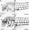
alg2p.G336*/p.G336* mutant embryos display severe alterations in vasculature anatomy. (A-B′) Homozygous alg2p.G336*/p.G336* allele in zFli::GFP reporter line (stage 40) showed an overall underdeveloped and thin vasculature anatomy (A) compared with unaffected stage-matched zFli::GFP siblings (B). Branching of cranial vessels (unfilled arrowheads) and developing gill vasculature were missing entirely in the homozygous mutant embryos. Main vessels in the yolk (y) were shortened and thin as were all vessels of the trunk and tail in mutants (A′) compared with control (B′). Note the very small atrium in mutants compared with controls (black arrowheads). CV, caudal vein; DA, dorsal aorta; DLAV, dorsal longitudinal anastomose vessel; ISV, intersegmental vessel. Scale bars: 200 µm.
Fig. 3.
Fig. 3.
Medaka Alg2:p.G336* and human ALG2:p.G347Vfs*26 are hypomorphic variants. (A,B) Lectin blots on total protein lysates of medaka alg2+/+ and homozygous alg2p.G336*/p.G336* mutant embryos (A) as well as human control and ALG2 CDG-patient fibroblasts (B). Intensity values normalized to internal loading control (Gapdh/β-actin) and wt or control samples. Con A, concanavalin A; WGA, wheat germ agglutinin. (C,D) xCGE-LIF-generated N-glycan fingerprints comparing medaka alg2+/+ (blue) with homozygous alg2p.G336*/p.G336* mutant embryos (red; C) and human control (blue) with ALG2 index patient (red; D) fibroblasts. N-glycan fingerprints represent average values from three biological replicates with the standard deviation shown as semi-transparent band. Asterisks indicate standard for migration time (MTU) normalization. Trends in observed quantitative changes for individual N-glycan structures are indicated by grey (complex-type) and green (high mannose-type) arrows with the direction indicating up- or downregulation. Peak numbers refer to identified N-glycan structures (see Table S2 for a list of all identified N-glycans). (E,F) Quantification of complex type (grey) and high mannose-type (green) N-glycan structures in medaka wt versus mutant (E) and control versus patient fibroblasts (F) from three biological replicates. Intensities represent the sum of all peak heights of complex- and high mannose-type N-glycan normalized to 100% of maximum for each chart, respectively.
Fig. 4.
Fig. 4.
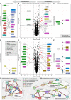
alg2p.G336*/p.G336* mutants show reduced levels of proteins involved in phototransduction but higher abundance of the glycosylation machinery. (A-F) Unbiased mass spectrometry of chemically labelled peptides derived from total protein lysates of deyolked wild-type (wt) alg2+/+ and alg2p.G336*/p.G336* whole hatchlings (A-C) and enucleated eyes (D-F) at stage 40. Differentially regulated proteins are shown in volcano plots (B-E), proteins exclusively detected in wild-type or mutants are presented in intensity plots (A,C,D,F). Coloured labelling and grouping according to (predicted) protein function retrieved by text-mining. (A,D) Intensity plots of proteins exclusively detected in wt samples amounted to 56 proteins in whole hatchling samples (A) and to seven proteins in the enucleated eye samples (D). (B,E) Volcano plots of protein abundance comparison between alg2+/+ and alg2p.G336*/p.G336* reveals no major regulation in protein abundance, yet distinct changes. Proteins with significant (P<0.05, dashed line) and more than 2-fold change (dotted lines) are indicated (red dots) and labelled. (C,F) Intensity plot of proteins exclusively detected in alg2p.G336*/p.G336* mutant samples amounted to three proteins for the whole hatchling samples (C) and 19 proteins in the enucleated eye samples (F). (G,H) Network analysis of functional associations and/or interactions of all regulated and exclusive proteins in eye samples. Different clusters of regulated proteins were identified using STRING software (medium confidence score, disconnected nodes hidden; Szklarczyk et al., 2019). Among the under-represented proteins in the mutants, photoreceptor-related players of the phototransduction pathway were enriched (G). Among the overrepresented ones, proteins involved in protein processing in the ER and of the nucleotide sugar metabolism pathways were pronounced (H). The coloured lines linking different proteins represent the types of evidence (blue: from curated databases; purple: experimentally determined; green: gene neighbourhood; black: co-expression; red: gene fusion; light green: text-mining). For full proteomics data, see Table S3. Asterisks in A and C indicate proteins associated with retinitis pigmentosa. Total protein extracts were derived from three replicates each comprising six stage 40, de-yolked hatchlings per genotype; analysis on eye samples were conducted on four replicates with 30 enucleated eyes of stage 40 hatchlings per genotype, respectively.
Fig. 5.
Fig. 5.
Rod photoreceptors are specifically affected in alg2p.G336*/p.G336* mutant retinae. (A-H) Time series of retinal development as depicted by DAPI and immunohistochemistry staining against rod (Rhodopsin) and cone (Zpr1) specific markers on cryotome sections in homozygous alg2p.G336*/p.G336* (A-D) and alg2+/+ (E-H) siblings at stage 32 (A,E), stage 35 (B,F) and hatching stage 40 (C-D,G-H). Separation of rod and cone photoreceptors from stage 35 onwards (F,G) failed in alg2p.G336*/p.G336* mutant embryos (B,C). The inner row of the alg2p.G336*/p.G336* ONL harbours pyknotic nuclei (C-D, white arrowheads) at the position of rod nuclei in wild-type embryos (G-H, black arrowheads). Zpr1 staining is unaffected in alg2p.G336*/p.G336* mutant embryos but appears irregular due to affected rod cells. CMZ, ciliary marginal zone; GCL, ganglion cell layer; INL, inner nuclear layer; IPL, inner plexiform layer; ONL, outer nuclear layer; OPL, outer plexiform layer. Insets show magnified views of the boxed areas in the panels above. Scale bars: 50 µm (main panels); 10 µm (insets). Six specimens per genotype and stage were used for analysis.
Fig. 6.
Fig. 6.
Sources of photoreceptor differentiation and regeneration are unaffected in alg2p.G336*/p.G336* hatchlings. Transverse cryotome sections through the eyes of stage 40 alg2p.G336*/p.G336* (A-A″) and alg2+/+ (B-B″) hatchlings. Immunohistochemistry detecting Rx2 (green) as a marker for retinal stem and progenitor cells, Müller glia (MG) cells and photoreceptors, and glutamine synthetase (GS, magenta) highlighting MG cell bodies did not reveal alterations between alg2p.G336*/p.G336* (A-A″) and wt controls (B-B″) in the CMZ nor the central retina except for the outer nuclear layer (insets). Here, Rx2 cone photoreceptor staining reconfirmed the monolayer of photoreceptors in alg2p.G336*/p.G336* hatchlings in contrast to the alg2+/+ bi-layer (compare with Fig. 5C,G). Scale bar: 50 µm. Insets show magnifications of boxed areas. Four specimens per genotype were used for analysis.
Fig. 7.
Fig. 7.
Hypo-N-glycosylation specifically triggers loss of rod photoreceptors in alg2p.G336*/p.G336* hatchlings. (A-F′) Transverse cryotome sections through the eyes of stage 32 (A,D), stage 35 (B,E) and hatching stage 40 (C,C′,F,F′) of alg2p.G336*/p.G336* (A-C′) and alg2+/+ (D-F′) hatchlings stained for apoptotic cells (TUNEL), cone photoreceptors (Zpr1) and nuclei (DAPI). Enrichment of TUNEL-positive cells in the outer nuclear layer of stage 40 hatchlings (ONL; white arrowheads) overlays with the location of rod photoreceptors (black arrowheads). (G) Quantification of TUNEL-positive cells on central eye sections. Highly significant enrichment of TUNEL-positive cells was observed in the ONL of alg2p.G336*/p.G336* hatchlings compared with stage-matched alg2+/+ siblings (P=0.009). All other layers and stages displayed slight but not significant enrichments of apoptotic cells in alg2p.G336*/p.G336* versus alg2+/+ samples (pairwise wilcoxon rank sum tests). Box plots depict median (thick black horizontal line); the extremes of the box are the 25th and 75th percentiles; the whiskers represent the minimum and maximum outliers; the filled circles represent extreme outliers. Non-filled scattered circles indicate raw data. CMZ, ciliary marginal zone; GCL, ganglion cell layer; INL, inner nuclear layer; IPL, inner plexiform layer; OPL, outer plexiform layer. Scale bars: 50 µm (main panels); 10 µm (insets). Eyes of six specimens per genotype and stage were used for analysis; non-central sections were excluded from analysis.
Fig. 8.
Fig. 8.
Human and medaka alg2 mRNA rescue the multisystemic phenotype in alg2p.G336*/p.G336*. (A) Individual genotyping of hatchlings at 4 days post hatching (dph) of batches descending from heterozygous alg2p.G336*/+ crosses revealed the absence of homozygous survivors. Rescue with medaka or human full-length alg2 mRNA restored their survival close to Mendelian distribution (see also Table S1). Number of hatchlings is shown at the base of the bars. (A′) The multisystemic phenotype of alg2p.G336*/p.G336* hatchlings were fully rescued by medaka alg2 mRNA but not GFP mRNA control injections at the one-cell stage. Exogenous supply of alg2 mRNA in alg2+/+ siblings did not result in any phenotype. Full-length human ALG2 mRNA rescued the gross morphology phenotype, but to a lesser extent, evident in the partially rescued snout size (brackets). (B) Alcian Blue staining (ventral view, stage 40) of GFP mRNA control (left) and full-length medaka alg2 mRNA rescue (central) injected at the one-cell stage into alg2p.G336*/p.G336* zygotes as well as full-length medaka alg2 mRNA injected into alg2+/+ (right) specimens. Cartilage anatomy in rescued and exogenous supplied alg2 in wt was indistinguishable. (C) Quantitative analysis of cartilage lengths, normalized to standard length (SL, distance between lenses; see Fig. 1F). Full-length alg2 mRNA completely rescues craniofacial cartilage structures in contrast to control GFP mRNA injections. Exogenous alg2 mRNA does not alter cartilage structures. m, Meckel's cartilage; pq, palatoquadrate; ch, ceratohyal. *P<0.05; ***P<0.001 (two-tailed nonparametric Student's t-test). n.s., not significant. Black lines indicate the mean. (D-E″) Immunohistochemistry staining against the photoreceptor-specific markers Rhodopsin (Rhod, rod photoreceptors, green) and Zpr1 (cone photoreceptors, magenta) and DAPI on transverse cryotome sections through the eyes of stage 40 alg2p.G336*/p.G336* and alg2+/+ hatchlings rescued after injection with full-length alg2 mRNA at the one-cell stage. Right-hand panels show magnifications of the boxed areas. Scale bars: 0.5 mm (A′,B); 50 µm (D″); 10 µm (right-hand panels in D-E″).

References

    1. Ba-Abbad, R., Leys, M., Wang, X., Chakarova, C., Waseem, N., Carss, K. J., Raymond, F. L., Bujakowska, K. M., Pierce, E. A., Mahroo, O. A.et al. (2018). Clinical features of a retinopathy associated with a dominant allele of the RGR gene. Invest. Ophthalmol. Vis. Sci. 59, 4812-4820. 10.1167/iovs.18-25061 - DOI - PMC - PubMed
    1. Boersema, P. J., Raijmakers, R., Lemeer, S., Mohammed, S. and Heck, A. J. R. (2009). Multiplex peptide stable isotope dimethyl labeling for quantitative proteomics. Nat. Protoc. 4, 484-494. 10.1038/nprot.2009.21 - DOI - PubMed
    1. Breitling, J. and Aebi, M. (2013). N-linked protein glycosylation in the endoplasmic reticulum. Cold Spring Harb. Perspect. Biol. 5, a013359. 10.1101/cshperspect.a013359 - DOI - PMC - PubMed
    1. Burns, D. M. and Touster, O. (1982). Purification and characterization of glucosidase II, an endoplasmic reticulum hydrolase involved in glycoprotein biosynthesis. J. Biol. Chem. 257, 9991-10000. 10.1016/S0021-9258(18)33975-9 - DOI - PubMed
    1. Cantagrel, V., Lefeber, D. J., Ng, B. G., Guan, Z., Silhavy, J. L., Bielas, S. L., Lehle, L., Hombauer, H., Adamowicz, M., Swiezewska, E.et al. (2010). SRD5A3 is required for converting polyprenol to dolichol and is mutated in a congenital glycosylation disorder. Cell 142, 203-217. 10.1016/j.cell.2010.06.001 - DOI - PMC - PubMed

Publication types