Review
. 2021 Aug;51(8):1911-1920.
doi: 10.1002/eji.202048994. Epub 2021 Jun 21.

Mechanism of action of PD-1 receptor/ligand targeted cancer immunotherapy

Affiliations
Review

Mechanism of action of PD-1 receptor/ligand targeted cancer immunotherapy

Jannie Borst et al. Eur J Immunol. 2021 Aug.

Abstract

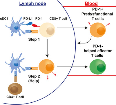
Immunotherapy targeting the Programmed Death (PD-1) receptor/ligand (L) "checkpoint" rapidly gains ground in the treatment of many cancer types. To increase treatment scope and efficacy, predictive biomarkers and rational selection of co-treatments are required. To meet these demands, we must understand PD-1 function in detail. We here outline recent insights into the regulation of the CD8+ T cell response by PD-1. The prevailing view has been that blockade of PD-1/ligand (L) interaction "reinvigorates" cytotoxic T lymphocytes (CTL) that were rendered dysfunctional in the tumor microenvironment (TME). However, this review stresses that tumors continuously communicate with adjacent draining lymph nodes (LNs) and that the PD-1 checkpoint also operates during T cell priming. We clarify the role of the PD-(L)1 system at the T cell/DC interface, where it regulates T cell receptor (TCR) signaling and CD28 costimulation and thus controls activation of tumor-specific T cells. We also highlight the importance of CD4+ T cell help during priming, which allows DCs to provide other costimulatory and cytokine signals required for optimal CTL differentiation and likely avoidance of a dysfunctional state. Therefore, we pose that PD-(L)1 blockade should exploit LN function and be combined with "help" signals to optimize CTL efficacy.

Keywords: PD-1; cytotoxic T cell; exhaustion; immunotherapy; priming.


Conflict of interest statement

The authors declare no commercial or financial conflict of interest.

Figures

Figure 1
Figure 1
Predysfunctional PD‐1+ CD8+ T cells in the TME. In the TME, a pool of PD‐1+ predysfunctional CD8+ T cells exists that undergoes self‐renewal. PD‐1 on the predysfunctional CD8+ T cells can interact with PD‐L1 on tumor cells or on cDCs. These interactions impair tumor cell killing, limiting tumor clearance and antigen availability. Furthermore, these interactions help drive the cells into an irreversibly exhausted state. Color codes: grey = tumor cell; blue = cDC; orange = predysfunctional CD8+ T cell; red = exhausted CD8+ T cell.
Figure 2
Figure 2
The PD‐1 checkpoint in T cell priming and effector stages. Upon killing of tumor cells, antigen can be taken up by cDCs and subsequently be transported via the lymph to tdLNs. Here, the cDCs prime naive T cells which leave the tdLN via the blood and enter the tumor. If these cells are properly differentiated into effector CTLs, they can kill more tumor cells, increasing antigen availability. However, interactions between PD‐1 and its ligands may limit efficacy of CTLs by limiting their priming and activity in the TME. Color codes: beige = naive CD8+ T cell; blue = cDC; orange = predysfunctional CD8+ T cell; grey = tumor cell.
Figure 3
Figure 3
Impact of PD‐L1 on TCR/CD28 signaling at the T cell/DC interface. (A) PD‐L1 expressed on activated DCs binds to PD‐1 on CD8+ T cells, thereby inhibiting the TCR/CD28 signal during priming. Co‐stimulation via CD28 is also impaired by the CTLA‐4 mediated downregulation of its ligand CD80. (B) When PD‐L1 forms a heterodimer with CD80 on the cell surface of the DC, signaling through PD‐1 is abrogated, but signaling through CD28 can still take place. Moreover, the CD80;PD‐L1 dimer can no longer be downregulated by CTLA‐4, resulting in an increase of CD28 co‐stimulation.
Figure 4
Figure 4
Proposed connection between help, optimal CTL priming and the PD‐1 checkpoint. Naive CD8+ T cells are activated by (migratory) cDC1s during the first step of priming. The PD‐1 axis inhibits TCR/CD28 signaling and thereby prevents clonal expansion. CD8+ T cells may leave the LN at this point, but are in an early stage of effector differentiation with limited cytotoxic and migratory abilities and expression of PD‐1 and other co‐inhibitory receptors. We propose that this “helpless” state equals the “predysfunctional” state identified in many studies. When appropriate activating signals are available, such as type I IFN, the CD8+ T cells undergo a second step of priming wherein help is delivered by activated CD4+ T cells via LN‐resident cDC1s. This leads to optimal effector differentiation of CD8+ T cells into potent CTLs that lack PD‐1 expression. Color codes: beige = naive CD8+ T cell; blue = cDC; orange = unhelped/predysfunctional CD8+ T cell; brown = CD4+ T helper cell; green = optimally primed/helped CTL.
Figure 5
Figure 5
Proposed effects of PD‐L1 targeted therapy in tdLNs and tumor. The effects of PD‐L1 blockade are threefold: (1) TCR/CD28 signaling is enabled during priming of naive (tumor‐specific) T cells, inducing proliferation of these cells. However, PD‐L1 blockade alone is not sufficient to allow the second step of priming in which help signals are delivered (dotted lines). Therefore, (2) PD‐L1 blockade in the tdLN increases the pool of (tumor‐specific) predysfunctional T cells. These cells will exit the tdLN and potentially reach the tumor, even though they have limited migratory ability. (3) In the TME, PD‐L1 blockade will also allow predysfunctional T cells to proliferate and transiently improve their cytotoxic capacities. However, ultimately these “reinvigorated” T cells will become exhausted. When “help mimicking” signals such as CD40 or CD27 agonism are added to PD‐L1 blockade, the second step of priming will be recapitulated and helped effector CTLs will be generated that lack PD‐1 expression and can more effectively kill the tumor cells. CD4+ T cell help (or its replacing signals) also allows for generation of CTL memory. Color codes: beige = naive CD8+ T cell; blue = cDC; orange = unhelped/predysfunctional CD8+ T cell; brown = CD4+ T helper cell; green = optimally primed/helped CTL; red = exhausted CD8+ T cell; grey = tumor cell.

References

    1. Sharma, P., Hu‐Lieskovan, S., Wargo, J. A. and Ribas, A., Primary, adaptive, and acquired resistance to cancer immunotherapy. Cell. 2017. 168: 707–723. - PMC - PubMed
    1. Ribas, A. and Wolchok, J. D., Cancer immunotherapy using checkpoint blockade. Science. 2018. 359: 1350–1355. - PMC - PubMed
    1. Schumacher, T. N. and Schreiber, R. D., Neoantigens in cancer immunotherapy. Science. 2015. 348: 69–74. - PubMed
    1. Palucka, A. K. and Coussens, L. M., The basis of oncoimmunology. Cell. 2016. 164: 1233–1247. - PMC - PubMed
    1. Topalian, S. L., Taube, J. M. and Pardoll, D. M., Neoadjuvant checkpoint blockade for cancer immunotherapy. Science. 2020. 367: eaax0182. - PMC - PubMed

Publication types

MeSH terms

Substances