

Gastrointestinal perforation secondary to COVID-19: Case reports and literature review
- PMID: 34106608
- PMCID: PMC8133225
- DOI: 10.1097/MD.0000000000025771
Gastrointestinal perforation secondary to COVID-19: Case reports and literature review
Abstract
Introduction: Corona virus disease-2019 (COVID-19) presents primarily with respiratory symptoms. However, extra respiratory manifestations are being frequently recognized including gastrointestinal involvement. The most common gastrointestinal symptoms are nausea, vomiting, diarrhoea and abdominal pain. Gastrointestinal perforation in association with COVID-19 is rarely reported in the literature.
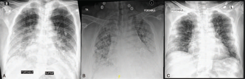
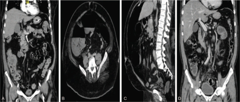
Patient concerns and diagnosis: In this series, we are reporting 3 cases with different presentations of gastrointestinal perforation in the setting of COVID-19. Two patients were admitted with critical COVID-19 pneumonia, both required intensive care, intubation and mechanical ventilation. The first one was an elderly gentleman who had difficult weaning from mechanical ventilation and required tracheostomy. During his stay in intensive care unit, he developed Candidemia without clear source. After transfer to the ward, he developed lower gastrointestinal bleeding and found by imaging to have sealed perforated cecal mass with radiological signs of peritonitis. The second one was an obese young gentleman who was found incidentally to have air under diaphragm. Computed tomography showed severe pneumoperitoneum with cecal and gastric wall perforation. The third case was an elderly gentleman who presented with severe COVID-19 pneumonia along with symptoms and signs of acute abdomen who was confirmed by imaging to have sigmoid diverticulitis with perforation and abscess collection.
Interventions: The first 2 cases were treated conservatively. The third one was treated surgically.
Outcome: Our cases had a variable hospital course but fortunately all were discharged in a good clinical condition.
Conclusion: Our aim from this series is to highlight this fatal complication to clinicians in order to enrich our understanding of this pandemic and as a result improve patients' outcome.
Copyright © 2021 the Author(s). Published by Wolters Kluwer Health, Inc.
Conflict of interest statement
The authors have no funding and conflicts of interests to disclose.
Figures
References
-
- https://www.who.int/news-room/detail/27-04-2020-who-timeline---covid-19 (Accessed September 23rd 2020).
Publication types
MeSH terms
LinkOut - more resources
Full Text Sources
Medical
