

T2-mapping increase is the prevalent imaging biomarker of myocardial involvement in active COVID-19: a Cardiovascular Magnetic Resonance study
- PMID: 34107985
- PMCID: PMC8189727
- DOI: 10.1186/s12968-021-00764-x
T2-mapping increase is the prevalent imaging biomarker of myocardial involvement in active COVID-19: a Cardiovascular Magnetic Resonance study
Abstract
Background: Early detection of myocardial involvement can be relevant in coronavirus disease 2019 (COVID-19) patients to timely target symptomatic treatment and decrease the occurrence of the cardiac sequelae of the infection. The aim of the present study was to assess the clinical value of cardiovascular magnetic resonance (CMR) in characterizing myocardial damage in active COVID-19 patients, through the correlation between qualitative and quantitative imaging biomarkers with clinical and laboratory evidence of myocardial injury.
Methods: In this retrospective observational cohort study, we enrolled 27 patients with diagnosis of active COVID-19 and suspected cardiac involvement, referred to our institution for CMR between March 2020 and January 2021. Clinical and laboratory characteristics, including high sensitivity troponin T (hs-cTnT), and CMR imaging data were obtained. Relationships between CMR parameters, clinical and laboratory findings were explored. Comparisons were made with age-, sex- and risk factor-matched control group of 27 individuals, including healthy controls and patients without other signs or history of myocardial disease, who underwent CMR examination between January 2020 and January 2021.
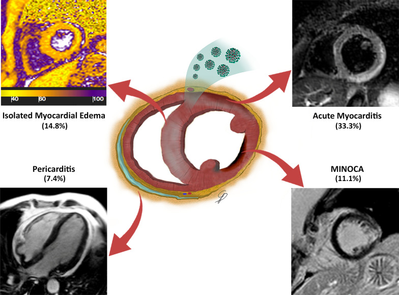
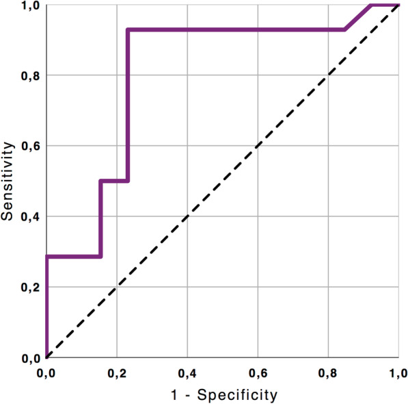
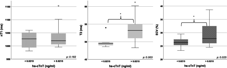
Results: The median (IQR) time interval between COVID-19 diagnosis and CMR examination was 20 (13.5-31.5) days. Hs-cTnT values were collected within 24 h prior to CMR and resulted abnormally increased in 18 patients (66.6%). A total of 20 cases (74%) presented tissue signal abnormalities, including increased myocardial native T1 (n = 11), myocardial T2 (n = 14) and extracellular volume fraction (ECV) (n = 10), late gadolinium enhancement (LGE) (n = 12) or pericardial enhancement (n = 2). A CMR diagnosis of myocarditis was established in 9 (33.3%), pericarditis in 2 (7.4%) and myocardial infarction with non-obstructive coronary arteries in 3 (11.11%) patients. T2 mapping values showed a moderate positive linear correlation with Hs-cTnT (r = 0.58; p = 0.002). A high degree positive linear correlation between ECV and Hs-cTnT was also found (r 0.77; p < 0.001).
Conclusions: CMR allows in vivo recognition and characterization of myocardial damage in a cohort of selected COVID-19 individuals by means of a multiparametric scanning protocol including conventional imaging and T1-T2 mapping sequences. Abnormal T2 mapping was the most commonly abnormality observed in our cohort and positively correlated with hs-cTnT values, reflecting the predominant edematous changes characterizing the active phase of disease.
Keywords: COVID-19; Cardiovascular Magnetic Resonance; Inflammation; Myocarditis; SARS-CoV-2; Troponin.
Conflict of interest statement
The authors declare that they have no competing interests.
Figures
References
-
- Catapano F, Marchitelli L, Cundari G, Cilia F, Mancuso G, Pambianchi G, et al. Role of advanced imaging in COVID-19 cardiovascular complications. Insights Imaging [Internet]. 2021 Feb 24 [cited 2021 Mar 3];12(1):28. http://www.ncbi.nlm.nih.gov/pubmed/33625637. - PMC - PubMed
-
- Alpert JS, Antman E, Apple F, Armstrong PW, Bassand JP, De Luna AB, et al. Myocardial infarction redefined—a consensus document of the Joint European Society of Cardiology/American College of Cardiology committee for the redefinition of myocardial infarction. Eur Heart J. 2000;21:1502–1513. doi: 10.1053/euhj.2000.2305. - DOI - PubMed
Publication types
MeSH terms
LinkOut - more resources
Full Text Sources
Medical
Research Materials
Miscellaneous
