. 2021 Jun 9;7(24):eabg5177.
doi: 10.1126/sciadv.abg5177. Print 2021 Jun.

Testicular germ cell-specific lncRNA, Teshl, is required for complete expression of Y chromosome genes and a normal offspring sex ratio

Affiliations

Testicular germ cell-specific lncRNA, Teshl, is required for complete expression of Y chromosome genes and a normal offspring sex ratio

Seong Hyeon Hong et al. Sci Adv. .

Abstract

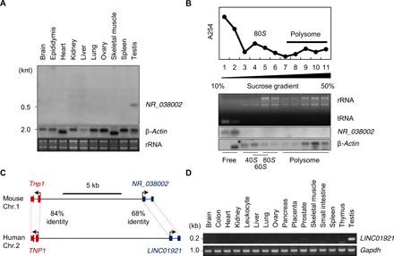
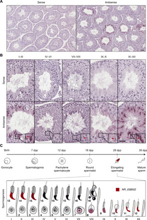
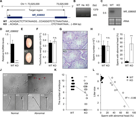
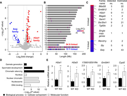
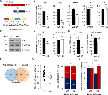
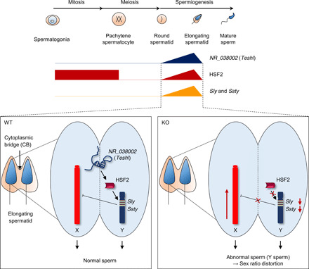
Heat shock factor 2 (HSF2) regulates the transcription of the male-specific region of the mouse Y chromosome long arm (MSYq) multicopy genes only in testes, but the molecular mechanism underlying this tissue specificity remains largely unknown. Here, we report that the testicular germ cell-specific long noncoding RNA (lncRNA), NR_038002, displays a characteristic spatiotemporal expression pattern in the nuclei of round and elongating spermatids. NR_038002-knockout male mice produced sperm with abnormal head morphology and exhibited reduced fertility accompanied by a female-biased sex ratio in offspring. Molecular analyses revealed that NR_038002 interacts with HSF2 and thereby activates expression of the MSYq genes. We designate NR_038002 as testicular germ cell-specific HSF2-interacting lncRNA (Teshl). Together, our study is the first to demonstrate that the testis specificity of HSF2 activity is regulated by the lncRNA Teshl and establishes a Teshl-HSF2-MSYq molecular axis for normal Y-bearing sperm qualities and consequent balanced offspring sex ratio.


Figures

Fig. 1
Fig. 1. Identification and characterization of the mouse testis–specific lncRNA, NR_038002.
(A) Tissue distribution of NR_038002 in various mouse tissues, as determined by Northern blotting. Middle and bottom panels represent a Northern blot of mouse β-actin mRNA and a denaturing agarose gel image of mouse 28S and 18S ribosomal RNA (rRNA), respectively (loading controls). (B) Polysome fractionation of NR_038002 lncRNA and β-actin mRNA in mouse testes followed by Northern blotting. Top graph: Absorbance at 254 nm for each fraction. Middle: Denaturing agarose gel electrophoresis of RNA extracted from each fraction. tRNA, transfer RNA. (C) Schematic representation of the genomic locus of NR_038002 and LINC01921 adjacent to Tnp1 in humans and mice, respectively. (D) Tissue distribution of LINC01921 in various human tissues, determined by reverse transcription polymerase chain reaction (RT-PCR).
Fig. 2
Fig. 2. Spatiotemporal expression patterns of NR_038002 in mouse testes.
(A) In situ hybridization analysis of NR_038002 in mouse testes using sense and antisense probes. Scale bar, 20 μm. (B) Higher magnification of in situ hybridization in serial sections for determination of expression stages and localization of NR_038002 in seminiferous tubules. NR_038002 expression was detected in nuclei of late round spermatids (weak signal) and elongating spermatids (strong signal). Note that the NR_038002 nuclear signal is different from hematoxylin and eosin (H&E) stain in color and shape (blurred), as indicated with red arrow in VII to VIII. Scale bar, 20 μm. (C) Diagram of 12-stage spermatogenesis represented by Roman numerals and 16-step spermiogenesis indicated by Arabic numbers in mice (53) showing the stages in which NR_038002 (red) is expressed.
Fig. 3
Fig. 3. Phenotypic analysis of NR_038002-KO male mice.
(A) Schematic diagram of the genomic locus of NR_038002 and the region deleted using CRISPR-Cas9 genome editing. The deleted region (−894 bp) of NR_038002 is indicated in the sequencing results. (B) Genotyping of NR_038002+/+ [wild-type (WT)], NR_038002+/− (He, heterozygote), and NR_038002−/− (KO) mice. (C) Northern blot of NR_038002 from WT and NR_038002-KO testes. (D) Quantitative RT-PCR (qRT-PCR) analyses of NR_038002 in WT and NR_038002-KO testes. Gapdh mRNA was used as a control. Data are presented a means ± SD (n = 4, ***P < 0.001, two-tailed Student’s t test). (E) Macroscopic appearance of adult testes from 8-week-old WT and NR_038002-KO mice. Scale bar, 0.2 mm. (F) Testis–to–body weight ratio in 8-week-old WT and NR_038002-KO mice (n = 6, n.s. P > 0.05, two-tailed Student’s t test). n.s., not significant. (G) H&E staining of seminiferous tubules of WT and NR_038002-KO mice. Scale bar, 40 μm. (H) Number of mature sperm from WT and NR_038002-KO mice (n = 6, n.s. P > 0.05, two-tailed Student’s t test). (I) Percentage of sperm with abnormal head morphology in WT and NR_038002-KO mice (n = 8, ***P < 0.001, two-tailed Student’s t test). (J) Representative spermatozoa from WT and NR_038002-KO mice. Red arrows indicate sperm with abnormal heads. (K) Number of embryos after mating with WT (n = 10) and NR_038002-KO (n = 8) male mice. Each dot represents an individual female mouse used for mating with WT (n = 28) or NR_038002-KO (n = 33) mice. Vaginal plugs were observed in all mated females. Data are presented a means ± SD (***P < 0.001, two-tailed Student’s t test). (L) Correlation between sperm with abnormal heads and litter size (n = 8). The dotted line shows the correlation between sperm head abnormality and litter size.
Fig. 4
Fig. 4. Loss of NR_038002 leads to aberrant up-regulation of X chromosomal genes.
(A) Volcano plots showing differentially expressed genes (DEGs) (fold change >1.5 and *P < 0.05) in WT and NR_038002-KO testes. (B) Distribution and heatmap of DEGs along the mouse chromosome, determined using ChromoMap. (C) Family names and detected and estimated numbers of multicopy sex-linked DEGs. (D) Gene Ontology enrichment analysis of DEGs. (E) qRT-PCR analysis of up-regulated X chromosomal genes. Gapdh mRNA was used as a control. Data are presented a means ± SD (n = 3, P > 0.05, *P < 0.05, and **P < 0.01, two-tailed Student’s t test).
Fig. 5
Fig. 5. Loss of NR_038002 leads to aberrant down-regulation of Y chromosomal multicopy genes and a female-biased sex ratio in offspring.
(A) Gene content and structure of mouse X and Y chromosomes. Representation of the ampliconic unit in the mouse Y chromosome, consisting of red, yellow, and blue core blocks. (B) qRT-PCR analysis of X and Y chromosomal multicopy genes in WT and NR_038002-KO mouse testes. Gapdh mRNA was used as a control. Data are presented as means ± SD (n = 3, n.s. P > 0.05, *P < 0.05, **P < 0.01, and ***P < 0.001, two-tailed Student’s t test). n.s., not significant. (C) Western blot of SLY and SLX/SLXL1 in WT and NR_038002-KO testes. (D) Densitometric analysis of SLY and SLX/SLXL1 in Western blots, normalized to α-tubulin. Data are presented as means ± SD (n = 3, n.s. P > 0.05, *P < 0.05, two-tailed Student’s t test). (E) qRT-PCR analysis of Sly and NR_038002 in WT and Sly–knockdown (KD) testes (n = 3, n.s. P > 0.05, **P < 0.01, two-tailed Student’s t test). (F) Venn diagram indicating overlapping DEGs in NR_038002-KO and Sly-KD testes. DEGs (fold change >1.5 and *P < 0.05) in sex chromosomes are indicated by the bold line. (G) Each dot represents the male pup ratio after mating with male WT (n = 18) and NR_038002-KO (n = 13) mice (*P < 0.05, two-tailed Student’s t test). (H) Bar graph showing the percentage of offspring of each sex, obtained by summation of the total number of pups (*P < 0.05, two-tailed Fisher’s exact t test). (I) Bar graph showing the average numbers of offspring of each sex, analyzed in (G) and (H). Data are presented as the means ± SEM (n.s. P > 0.05, *P < 0.05, two-tailed Student’s t test).
Fig. 6
Fig. 6. NR_038002 interacts with HSF2 to activate Y chromosomal multicopy genes.
(A) qRT-PCR analysis of Hsf1 and Hsf2 in WT and NR_038002-KO testes. Gapdh mRNA was used as a control. Data are presented as means ± SD (n = 3, n.s. P > 0.05, two-tailed Student’s t test). n.s., not significant. (B) Western blot of HSF1 and HSF2 in WT and NR_038002-KO testes. (C) Densitometric analysis of HSF1 and HSF2 in Western blots, normalized to α-tubulin. Data are presented as means ± SD (n = 3, n.s. P > 0.05, two-tailed Student’s t test). (D) Schematic diagram of RNA pull-down assay. (E) Western blot of eluates obtained by RNA pull-down using specific anti-HSF1 and anti-HSF2 antibodies. Bottom panel shows biotinylated full-length antisense and sense strands of NR_038002. (F) Western blot of eluates obtained by RNA pull-down using the anti-HSF2 antibody. Bottom panel shows sense strands of biotinylated full-length and fragmented [1 to 100 nt, 101 to 30 nt, and 231 to 416 nt] NR_038002. (G) RNA immunoprecipitation (RIP) PCR analysis of NR_038002, which binds to HSF2 in mouse testes. Gapdh mRNA, which does not bind to HSF2, served as the negative control. IgG, immunoglobulin G. (H) RIP-qPCR analysis of NR_038002. Data are presented as means ± SD (n = 3, *P < 0.05, two-tailed Student’s t test). (I) Chromatin immunoprecipitation qPCR analysis of Sly, Ssty1, and Ssty2 promoter regions, which bind HSF2, in WT and NR_038002-KO mouse testes. The intergenic region, which does not bind HSF2, served as a negative control. Data are presented as means ± SD (n = 3, n.s. P > 0.05, *P < 0.05, two-tailed Student’s t test). (J) Model of NR_038002 function, showing that NR_038002 binding to HSF2 activates multicopy Y chromosomal Sly and Ssty genes.
Fig. 7
Fig. 7. Schematic diagram showing production of normal Y-bearing sperm by NR_038002 (Teshl)–dependent HSF2 action on the male-specific Y chromosome long arm.
NR_038002 (Teshl) exhibits spatiotemporally expression patterns in the nuclei of elongating spermatids in WT mouse testes. HSF2 is expressed during early spermatogenesis, inactivated during meiosis, and reactivated beginning in round spermatids. Teshl plays an important role in activating the Y chromosomal multicopy genes Sly and Ssty by directing the binding of HSF2 to target regions, which results in down-regulation of X chromosome genes. In Teshl-KO mouse testes, HSF2 cannot bind to the promoter regions of Sly and Ssty, resulting in a significant decrease in their expression levels and consequent up-regulation of spermatid-expressed genes located on the X chromosome. Therefore, Y-bearing sperm with abnormal head morphology are produced, and offspring exhibit a female-biased sex ratio.

References

    1. Kapranov P., Cheng J., Dike S., Nix D. A., Duttagupta R., Willingham A. T., Stadler P. F., Hertel J., Hackermuller J., Hofacker I. L., Bell I., Cheung E., Drenkow J., Dumais E., Patel S., Helt G., Ganesh M., Ghosh S., Piccolboni A., Sementchenko V., Tammana H., Gingeras T. R., RNA maps reveal new RNA classes and a possible function for pervasive transcription. Science 316, 1484–1488 (2007). - PubMed
    1. Fatica A., Bozzoni I., Long non-coding RNAs: New players in cell differentiation and development. Nat. Rev. Genet. 15, 7–21 (2014). - PubMed
    1. Long Y., Wang X., Youmans D. T., Cech T. R., How do lncRNAs regulate transcription? Sci. Adv. 3, eaao2110 (2017). - PMC - PubMed
    1. Kutter C., Watt S., Stefflova K., Wilson M. D., Goncalves A., Ponting C. P., Odom D. T., Marques A. C., Rapid turnover of long noncoding RNAs and the evolution of gene expression. PLOS Genet. 8, e1002841 (2012). - PMC - PubMed
    1. Necsulea A., Soumillon M., Warnefors M., Liechti A., Daish T., Zeller U., Baker J. C., Grutzner F., Kaessmann H., The evolution of lncRNA repertoires and expression patterns in tetrapods. Nature 505, 635–640 (2014). - PubMed

Publication types