. 2021 Mar 1;45(2):75-86.
doi: 10.1080/01658107.2020.1856143. eCollection 2021.

The Effect of Congenital and Acquired Bilateral Anophthalmia on Brain Structure

Affiliations

The Effect of Congenital and Acquired Bilateral Anophthalmia on Brain Structure

Holly Bridge et al. Neuroophthalmology. .

Abstract

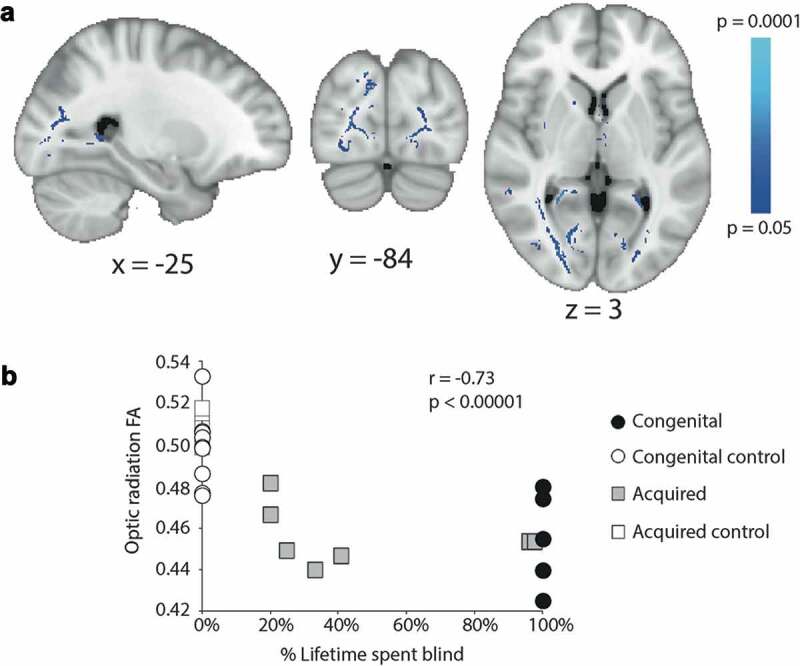
The aim of this study was to compare the pattern of changes in brain structure resulting from congenital and acquired bilateral anophthalmia. Brain structure was investigated using 3T magnetic resonance imaging (MRI) in Oxford (congenital) or Manchester (acquired). T1-weighted structural and diffusion-weighted scans were acquired from people with anophthalmia and sighted control participants. Differences in grey matter between the groups were quantified using voxel-based morphometry and differences in white matter microstructure using tract-based spatial statistics. Quantification of optic nerve volume and cortical thickness in visual regions was also performed in all groups. The optic nerve was reduced in volume in both anophthalmic populations, but to a greater extent in the congenital group and anophthalmia acquired at an early age. A similar pattern was found for the white matter microstructure throughout the occipitotemporal regions of the brain, suggesting a greater reduction of integrity with increasing duration of anophthalmia. In contrast, grey matter volume changes differed between the two groups, with the acquired anophthalmia group showing a decrease in the calcarine sulcus, corresponding to the area that would have been peripheral primary visual cortex. In contrast, the acquired anophthalmia group showed a decrease in grey matter volume in the calcarine sulcus corresponding to the area that would have been peripheral primary visual cortex. There are both qualitative and quantitative differences in structural brain changes in congenital and acquired anophthalmia, indicating differential effects of development and degeneration.

Keywords: Anophthalmia; cortical thickness; diffusion imaging; grey matter; magnetic resonance imaging; optic nerve.


Figures

Figure 1.
Figure 1.
Optic nerve analysis using T1-weighted MPRAGE images. Axial slices show the expected location of the optic nerve in all congenital (a) and six of the acquired (b) anophthalmic cases. For comparison, the optic nerves from two example controls are also shown. In each case, white arrows point to the optic nerve if present. Mean optic nerve volume for each group (c) is plotted. Error bars represent standard error of the mean. Finally, optic nerve volume for each acquired anophthalmic case and relevant controls was plotted against % life spent blind, revealing a significant negative correlation (d, Pearson’s r = 0.56, p < .005)
Figure 2.
Figure 2.
Reduced grey matter volume in acquired anophthalmia compared to sighted controls in the anterior calcarine in the right hemisphere. The colour represents the significance of the difference between the two groups. Data are shown on the MNI standard brain
Figure 3.
Figure 3.
Cortical thickness in visual regions of interest. A shows the cortical thickness measures in the congenital anophthalmia group and controls. Cortical thickness in V1 and V2 of the congenital anophthalmia groups was significantly greater than sighted controls. B shows the same data for the acquired anophthalmia group. There are no significant differences in cortical thickness in any areas in the acquired anophthalmia group. C shows the correlation between % life spent blind and V1 cortical thickness. D shows the correlation between age of blindness onset and V1 cortical thickness. Asterisks indicate p < .005
Figure 4.
Figure 4.
Reduced FA in congenital (a) and acquired (b) anophthalmia groups compared to sighted controls. Red-yellow regions indicate significantly lower FA in the congenital anophthalmia group and blue shows those significantly lower in the acquired anophthalmia group. For visualisation purposes, all statistical maps are thresholded at p < .05 (after TFCE correction for multiple comparisons)
Figure 5.
Figure 5.
A shows the white matter in which the reduction in FA correlates inversely with the percentage of life spent blind across the acquired anophthalmia group. Mean optic radiation FA (both hemispheres) plotted against percentage of lifetime spent blind for the congenital anophthalmics (black circles, all 100%), acquired anophthalmics (grey squares), congenital controls (white circles, all 0%) and acquired controls (white squares, all 0%)

References

    1. Noppeney U, Friston KJ, Ashburner J, Frackowiak R, Price CJ.. Early visual deprivation induces structural plasticity in gray and white matter. Curr Biol. 2005;15(13):R488–490. doi:10.1016/j.cub.2005.06.053. - DOI - PubMed
    1. Ptito M, Schneider FC, Paulson OB, Kupers R. Alterations of the visual pathways in congenital blindness. Exp Brain Res. 2008;187(1):41–49. doi:10.1007/s00221-008-1273-4. - DOI - PubMed
    1. Bock AS, Saenz M, Tungaraza R, Boynton GM, Bridge H, Fine I. Visual callosal topography in the absence of retinal input. NeuroImage. 2013;81:325–334. doi:10.1016/j.neuroimage.2013.05.038. - DOI - PMC - PubMed
    1. Aguirre GK, Datta R, Benson NC, et al. Patterns of individual variation in visual pathway structure and function in the sighted and blind. PLoS One. 2016;11(11):e0164677. doi:10.1371/journal.pone.0164677. - DOI - PMC - PubMed
    1. Good CD, Johnsrude IS, Ashburner J, Henson RN, Friston KJ, Frackowiak RS. A voxel-based morphometric study of ageing in 465 normal adult human brains. NeuroImage. 2001;14:21–36. doi:10.1006/nimg.2001.0786. - DOI - PubMed

LinkOut - more resources