

Biomechanics of the Distal Radioulnar Joint During In Vivo Forearm Pronosupination
- PMID: 34109063
- PMCID: PMC8169167
- DOI: 10.1055/s-0040-1722334
Biomechanics of the Distal Radioulnar Joint During In Vivo Forearm Pronosupination
Abstract
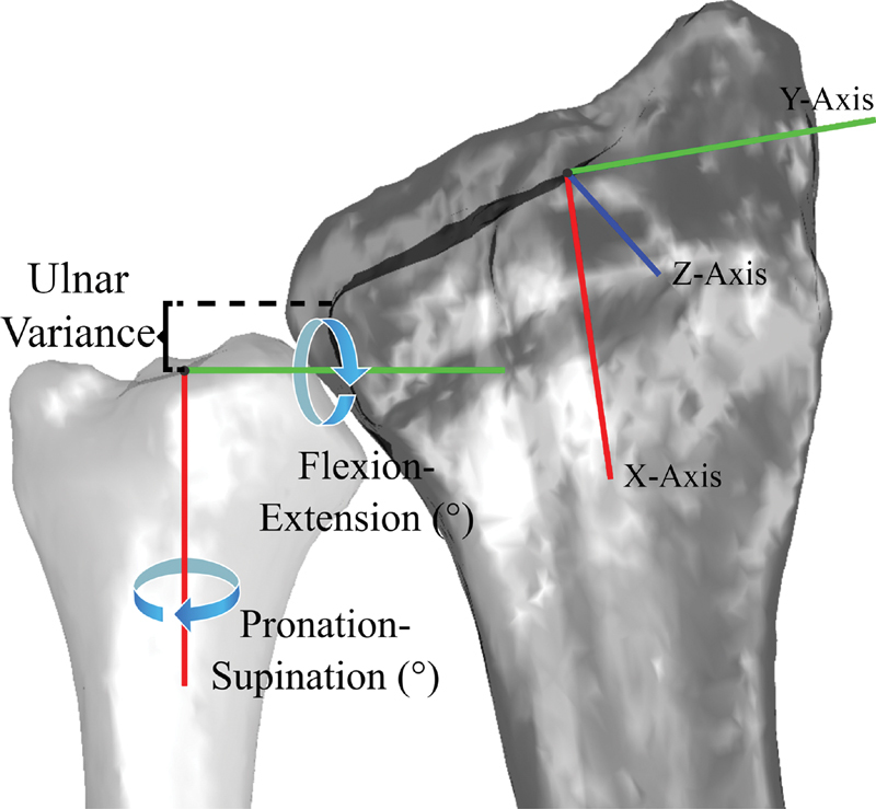
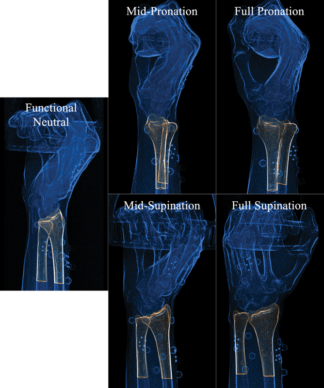
Background Ulnar variance (UV) and center of rotation (COR) location at the level of the distal radioulnar joint (DRUJ) change with forearm rotation. Nevertheless, these parameters have not been assessed dynamically during active in vivo pronosupination. This assessment could help us to improve our diagnosis and treatment strategies. Questions/purposes We sought to (1) mathematically model the UV change, and (2) determine the dynamic COR's location during active pronosupination. Methods We used biplanar videoradiography to study DRUJ during in vivo pronation and supination in nine healthy subjects. UV was defined as the proximal-distal distance of ulnar fovea with respect to the radial sigmoid notch, and COR was calculated using helical axis of motion parameters. The continuous change of UV was evaluated using a generalized linear regression model. Results A second-degree polynomial with R 2 of 0.85 was able to model the UV changes. Maximum negative UV occurred at 38.0 degrees supination and maximum positive UV occurred at maximum pronation. At maximum pronation, the COR was located 0.5 ± 1.8 mm ulnarly and 0.6 ± 0.8 mm volarly from the center of the ulnar fovea, while at maximum supination, the COR was located 0.2 ± 0.6 mm radially and 2.0 ± 0.5 mm volarly. Conclusion Changes in UV and volar translation of the COR are nonlinear at the DRUJ during pronosupination. Clinical Relevance Understanding the dynamic nature of UV as a function of pronosupination can help guide accurate evaluation and treatment of wrist pathology where the UV is an important consideration. The dynamic behavior of COR might be useful in designing DRUJ replacement implants to match the anatomical motion.
Keywords: DRUJ; center of rotation; kinematics; radioulnar joint; ulnar variance.
Thieme. All rights reserved.
Conflict of interest statement
Conflict of Interest None declared.
Figures


References
-
- Linscheid R L. Biomechanics of the distal radioulnar joint. Clin Orthop Relat Res. 1992;(275):46–55. - PubMed
-
- Shaaban H, Giakas G, Bolton M, Williams R, Scheker L R, Lees V C. The distal radioulnar joint as a load-bearing mechanism—a biomechanical study. J Hand Surg Am. 2004;29(01):85–95. - PubMed
-
- Epner R A, Bowers W H, Guilford W B. Ulnar variance—the effect of wrist positioning and roentgen filming technique. J Hand Surg Am. 1982;7(03):298–305. - PubMed
-
- Friedman S L, Palmer A K. The ulnar impaction syndrome. Hand Clin. 1991;7(02):295–310. - PubMed
-
- Laino D K, Petchprapa C N, Lee S K. Ulnar variance: correlation of plain radiographs, computed tomography, and magnetic resonance imaging with anatomic dissection. J Hand Surg Am. 2012;37(01):90–97. - PubMed
