

Label-Free Macroscopic Fluorescence Lifetime Imaging of Brain Tumors
- PMID: 34109119
- PMCID: PMC8181388
- DOI: 10.3389/fonc.2021.666059
Label-Free Macroscopic Fluorescence Lifetime Imaging of Brain Tumors
Abstract
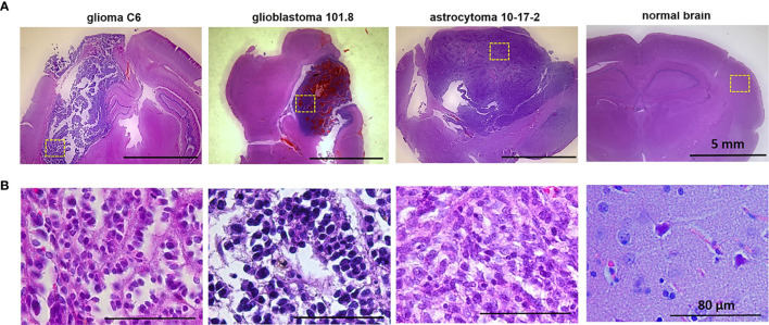
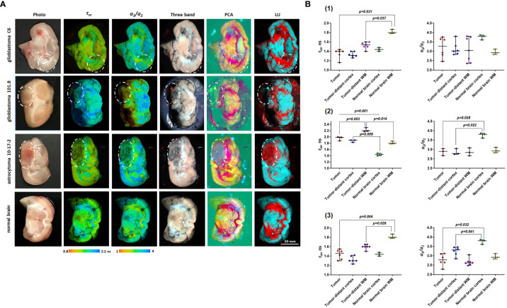
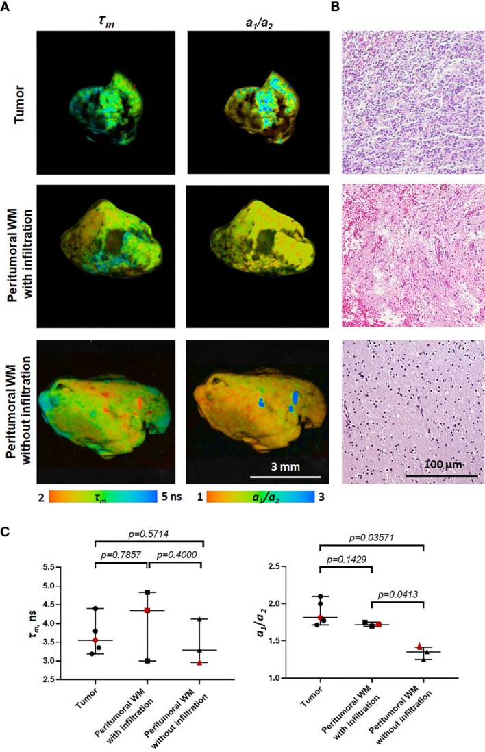
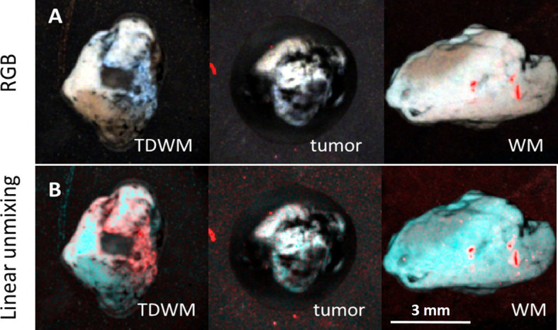
Advanced stage glioma is the most aggressive form of malignant brain tumors with a short survival time. Real-time pathology assisted, or image guided surgical procedures that eliminate tumors promise to improve the clinical outcome and prolong the lives of patients. Our work is focused on the development of a rapid and sensitive assay for intraoperative diagnostics of glioma and identification of optical markers essential for differentiation between tumors and healthy brain tissues. We utilized fluorescence lifetime imaging (FLIM) of endogenous fluorophores related to metabolism of the glioma from freshly excised brains tissues. Macroscopic time-resolved fluorescence images of three intracranial animal glioma models and surgical samples of patients' glioblastoma together with the white matter have been collected. Several established and new algorithms were applied to identify the imaging markers of the tumors. We found that fluorescence lifetime parameters characteristic of the glioma provided background for differentiation between the tumors and intact brain tissues. All three rat tumor models demonstrated substantial differences between the malignant and normal tissue. Similarly, tumors from patients demonstrated statistically significant differences from the peritumoral white matter without infiltration. While the data and the analysis presented in this paper are preliminary and further investigation with a larger number of samples is required, the proposed approach based on the macroscopic FLIM has a high potential for diagnostics of glioma and evaluation of the surgical margins of gliomas.
Keywords: FLIM; autofluorescence; fluorescence lifetime imaging; glioblastoma; image processing; rat glioma model.
Copyright © 2021 Lukina, Yashin, Kiseleva, Alekseeva, Dudenkova, Zagaynova, Bederina, Medyanic, Becker, Mishra, Berezin, Shcheslavskiy and Shirmanova.
Conflict of interest statement
MB is the founder of HSpeQ LLC, a hyperspectral imaging company that develops IDCube software. WB and VS was employed by the company Becker&Hickl GmbH. The remaining authors declare that the research was conducted in the absence of any commercial or financial relationships that could be construed as a potential conflict of interest.
Figures
Similar articles
-
First in patient assessment of brain tumor infiltrative margins using simultaneous time-resolved measurements of 5-ALA-induced PpIX fluorescence and tissue autofluorescence.J Biomed Opt. 2022 Feb;27(2):020501. doi: 10.1117/1.JBO.27.2.020501. J Biomed Opt. 2022. PMID: 35112514 Free PMC article.
-
Delineation of gastrointestinal tumors biopsies using a fluorescence lifetime imaging optical fiber probe.J Biophotonics. 2024 Jul 16:e202400122. doi: 10.1002/jbio.202400122. Online ahead of print. J Biophotonics. 2024. PMID: 39014559
-
Multiphoton excitation of autofluorescence for microscopy of glioma tissue.Neurosurgery. 2006 Apr;58(4):759-67; discussion 759-67. doi: 10.1227/01.NEU.0000204885.45644.22. Neurosurgery. 2006. PMID: 16575340
-
Mesoscopic fluorescence lifetime imaging: Fundamental principles, clinical applications and future directions.J Biophotonics. 2021 Jun;14(6):e202000472. doi: 10.1002/jbio.202000472. Epub 2021 Mar 29. J Biophotonics. 2021. PMID: 33710785 Free PMC article. Review.
-
Fluorescence lifetime imaging--techniques and applications.J Microsc. 2012 Aug;247(2):119-36. doi: 10.1111/j.1365-2818.2012.03618.x. Epub 2012 May 24. J Microsc. 2012. PMID: 22621335 Review.
Cited by
-
Flavin fluorescence lifetime and autofluorescence optical redox ratio for improved visualization and classification of brain tumors.Front Oncol. 2023 Feb 20;13:1105648. doi: 10.3389/fonc.2023.1105648. eCollection 2023. Front Oncol. 2023. PMID: 36890834 Free PMC article.
-
Near-Infrared Inorganic Nanomaterials for Precise Diagnosis and Therapy.Front Bioeng Biotechnol. 2021 Oct 26;9:768927. doi: 10.3389/fbioe.2021.768927. eCollection 2021. Front Bioeng Biotechnol. 2021. PMID: 34765596 Free PMC article. Review.
-
In vivo characterization of the human glioblastoma infiltrative edge with label-free intraoperative fluorescence lifetime imaging.Biomed Opt Express. 2023 Apr 24;14(5):2196-2208. doi: 10.1364/BOE.481304. eCollection 2023 May 1. Biomed Opt Express. 2023. PMID: 37206147 Free PMC article.
-
OCT-Guided Surgery for Gliomas: Current Concept and Future Perspectives.Diagnostics (Basel). 2022 Jan 28;12(2):335. doi: 10.3390/diagnostics12020335. Diagnostics (Basel). 2022. PMID: 35204427 Free PMC article. Review.
-
Raman Imaging and Fluorescence Lifetime Imaging Microscopy for Diagnosis of Cancer State and Metabolic Monitoring.Cancers (Basel). 2021 Nov 13;13(22):5682. doi: 10.3390/cancers13225682. Cancers (Basel). 2021. PMID: 34830837 Free PMC article. Review.
References
-
- Erkkilä MT, Reichert D, Gesperger J, Kiesel B, Roetzer T, Mercea PA, et al. . Macroscopic Fluorescence-Lifetime Imaging of NADH and Protoporphyrin IX Improves the Detection and Grading of 5-Aminolevulinic Acid-Stained Brain Tumors. Sci Rep (2020) 10:20492. 10.1038/s41598-020-77268-8 - DOI - PMC - PubMed
Grants and funding
LinkOut - more resources
Full Text Sources