High Resolution View on the Regulation of Recombinase Accumulation in Mammalian Meiosis
- PMID: 34109178
- PMCID: PMC8181746
- DOI: 10.3389/fcell.2021.672191
High Resolution View on the Regulation of Recombinase Accumulation in Mammalian Meiosis
Abstract
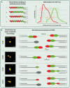
A distinguishing feature of meiotic DNA double-strand breaks (DSBs), compared to DSBs in somatic cells, is the fact that they are induced in a programmed and specifically orchestrated manner, which includes chromatin remodeling prior to DSB induction. In addition, the meiotic homologous recombination (HR) repair process that follows, is different from HR repair of accidental DSBs in somatic cells. For instance, meiotic HR involves preferred use of the homolog instead of the sister chromatid as a repair template and subsequent formation of crossovers and non-crossovers in a tightly regulated manner. An important outcome of this distinct repair pathway is the pairing of homologous chromosomes. Central to the initial steps in homology recognition during meiotic HR is the cooperation between the strand exchange proteins (recombinases) RAD51 and its meiosis-specific paralog DMC1. Despite our understanding of their enzymatic activity, details on the regulation of their assembly and subsequent molecular organization at meiotic DSBs in mammals have remained largely enigmatic. In this review, we summarize recent mouse data on recombinase regulation via meiosis-specific factors. Also, we reflect on bulk "omics" studies of initial meiotic DSB processing, compare these with studies using super-resolution microscopy in single cells, at single DSB sites, and explore the implications of these findings for our understanding of the molecular mechanisms underlying meiotic HR regulation.
Keywords: ChIP-seq; DSB repair; meiosis; recombinase; super-resolution microscopy.
Copyright © 2021 Mhaskar, Koornneef, Zelensky, Houtsmuller and Baarends.
Conflict of interest statement
The authors declare that the research was conducted in the absence of any commercial or financial relationships that could be construed as a potential conflict of interest.
Figures


Similar articles
-
Pseudosynapsis and decreased stringency of meiotic repair pathway choice on the hemizygous sex chromosome of Caenorhabditis elegans males.Genetics. 2014 Jun;197(2):543-60. doi: 10.1534/genetics.114.164152. Genetics. 2014. PMID: 24939994 Free PMC article.
-
Switching yeast from meiosis to mitosis: double-strand break repair, recombination and synaptonemal complex.Genes Cells. 1997 Aug;2(8):487-98. doi: 10.1046/j.1365-2443.1997.1370335.x. Genes Cells. 1997. PMID: 9348039
-
A meiosis-specific BRCA2 binding protein recruits recombinases to DNA double-strand breaks to ensure homologous recombination.Nat Commun. 2019 Feb 13;10(1):722. doi: 10.1038/s41467-019-08676-2. Nat Commun. 2019. PMID: 30760716 Free PMC article.
-
Biochemical attributes of mitotic and meiotic presynaptic complexes.DNA Repair (Amst). 2018 Nov;71:148-157. doi: 10.1016/j.dnarep.2018.08.018. Epub 2018 Aug 23. DNA Repair (Amst). 2018. PMID: 30195641 Free PMC article. Review.
-
The meiotic-specific Mek1 kinase in budding yeast regulates interhomolog recombination and coordinates meiotic progression with double-strand break repair.Curr Genet. 2019 Jun;65(3):631-641. doi: 10.1007/s00294-019-00937-3. Epub 2019 Jan 22. Curr Genet. 2019. PMID: 30671596 Free PMC article. Review.
Cited by
-
Methods of Detection and Mechanisms of Origin of Complex Structural Genome Variations.Methods Mol Biol. 2024;2825:39-65. doi: 10.1007/978-1-0716-3946-7_2. Methods Mol Biol. 2024. PMID: 38913302 Review.
-
Single-Cell RNA Sequencing of the Testis of Ciona intestinalis Reveals the Dynamic Transcriptional Profile of Spermatogenesis in Protochordates.Cells. 2022 Dec 8;11(24):3978. doi: 10.3390/cells11243978. Cells. 2022. PMID: 36552742 Free PMC article.
-
Multi-color dSTORM microscopy in Hormad1-/- spermatocytes reveals alterations in meiotic recombination intermediates and synaptonemal complex structure.PLoS Genet. 2022 Jul 20;18(7):e1010046. doi: 10.1371/journal.pgen.1010046. eCollection 2022 Jul. PLoS Genet. 2022. PMID: 35857787 Free PMC article.
-
RNase H1 facilitates recombinase recruitment by degrading DNA-RNA hybrids during meiosis.Nucleic Acids Res. 2023 Aug 11;51(14):7357-7375. doi: 10.1093/nar/gkad524. Nucleic Acids Res. 2023. PMID: 37378420 Free PMC article.
-
Single-Cell Atlas of Adult Testis in Protogynous Hermaphroditic Orange-Spotted Grouper, Epinephelus coioides.Int J Mol Sci. 2021 Nov 22;22(22):12607. doi: 10.3390/ijms222212607. Int J Mol Sci. 2021. PMID: 34830486 Free PMC article.
References
Publication types
LinkOut - more resources
Full Text Sources
Research Materials

