. 2022 Feb;71(2):237-249.
doi: 10.1007/s00262-021-02976-7. Epub 2021 Jun 10.

T cells, particularly activated CD4+ cells, maintain anti-CD20-mediated NK cell viability and antibody dependent cellular cytotoxicity

Affiliations

T cells, particularly activated CD4+ cells, maintain anti-CD20-mediated NK cell viability and antibody dependent cellular cytotoxicity

Zhaoming Wang et al. Cancer Immunol Immunother. 2022 Feb.

Abstract

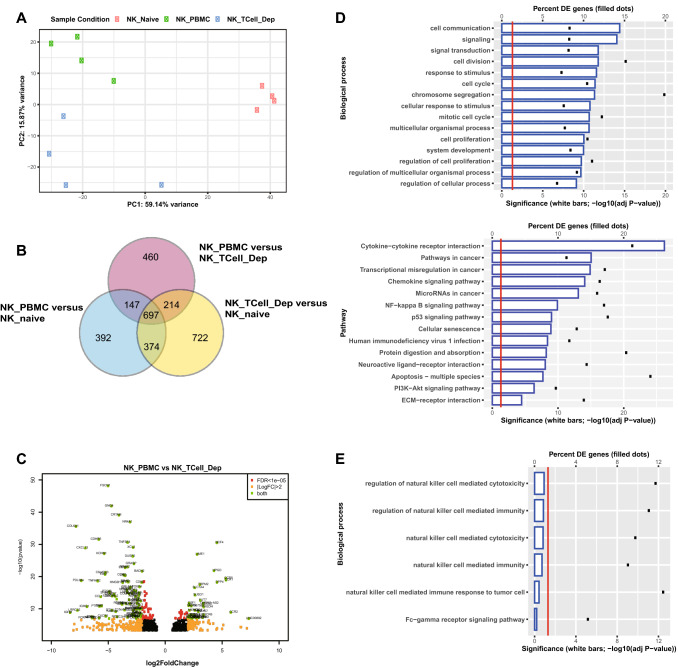
Anti-CD20 monoclonal antibody (mAb) therapy is a mainstay of therapy for B cell malignancies, however many patients fail to respond or eventually develop resistance. The current understanding of mechanisms responsible for this resistance is limited. When peripheral blood mononuclear cells of healthy donors were cultured with Raji cells for 7 days, rituximab (RTX) induced NK cell-mediated antibody-dependent cellular cytotoxicity (ADCC), enhanced NK cell viability and increased or maintained NK expression of CD56, CD16, CD57 and KIR. T cells, mainly CD4+, mediated these changes in a contact-dependent manner, with local T cell production of IL2 playing a central role. Similar findings were found when autologous B cells were used as target cells demonstrating the need for T cell help was not due to allogenic reaction. Results with other anti-CD20 and anti-EGFR antibodies were consistent. Small numbers of T cells activated by anti-CD3/CD28 beads or bispecific antibody enhanced RTX-mediated NK cell ADCC, viability and phenotypical changes. Pathway analysis of bulk NK cell mRNA sequencing after activation by RTX with and without T cells was consistent with T cells maintaining the viability of the activated NK cells. These findings suggest T cell help, mediated in large part by local production of IL2, contributes to NK cell ADCC and viability, and that activating T cells in the tumor microenvironment, such as through the use of anti-CD3 based bispecific antibodies, could enhance the efficacy of anti-CD20 and other mAb therapies where NK-mediated ADCC is a primary mechanism of action.

Keywords: ADCC; Bispecific antibody; NK cell; Rituximab; T cell.


Figures

Fig. 1
Fig. 1
Long-term exposure to RTX impacts NK cell function and phenotype. PBMC were cocultured with Raji cells and RTX or TRA. NK cell function and phenotype were examined at different time points. a CD19+ target cells are progressively eliminated over time in response to RTX. b RTX maintains the number of NK cells within PBMCs. c RTX induces proliferation of NK cells within PBMC at 7 days as determined by CFSE dilution. d, e RTX progressively increases the percent of CD56bright NK cells beginning at day 3. n = 6. Cell counts at 0 h were used to normalize cell numbers. f Patient 4 has circulating tumor cells, while the others have no detectable circulating tumor cells. g The fraction of CD56bright NK cells increased following RTX in the patient with circulating malignant cells but not in patients without circulating malignant cells
Fig. 2
Fig. 2
CD56dim NK cells transit into CD56bright NK cells in response to long-term RTX activation. a Isolated CD56dim and CD56bright NK cells were stained with CFSE and CellTracker Red, respectively. CFSE-labeled CD56dim and Red-labeled CD56bright NK were added to autologous PBMC and cocultured with Raji cells and RTX or TRA. b As illustrated by a day 7 histogram gated on CFSE-labeled CD56dim NK cells, CFSE-labeled CD56dim NK cells proliferate and adopt a CD56bright phenotype in the RTX group. n = 3. c The number of CellTracker Red labeled-CD56bright NK cells does not increase in response to RTX. n = 3. d–i CD16, CD57 and KIR were largely expressed by resting CD56dim NK cells. RTX-induced CD56bright NK cells express high levels of CD16, CD57 and KIR. n = 6. Cell counts at 0 h were used to normalize cell numbers
Fig. 3
Fig. 3
T cells are required for RTX-mediated NK cell cytotoxicity, viability, CD16 re-expression and CD56dim to CD56bright transition. Unfractionated PBMC or PBMC depleted of CD3+, CD4+, or CD8+ cells were cocultured with Raji cells and RTX or TRA. a, b CD19+ target cells are mostly eliminated by RTX on day 7. RTX-mediated elimination of CD19+ cells is inhibited by the depletion of CD3+ T cells. c The number of NK cells remaining in intact PBMCs is maintained by RTX but not by TRA. However, NK cell numbers are not maintained after CD3+ T cell depletion. d, e The expression of CD16 is downregulated on NK cells by RTX activation at 20 h and recovers on day 7. CD16 re-expression is not observed after CD3+ T cell depletion. f, g CD56dim to CD56bright NK transition is induced in unfractionated PBMC after 7 days. This transition is inhibited by depletion of CD3+ or CD4+ cells but not after depletion of CD8+ cells. Cell counts in the TRA + PBMC group were used to normalize cell numbers. n = 4–6. Dep: depleted
Fig. 4
Fig. 4
T cells are required for RTX-mediated NK cell responses in the autologous system. Unfractionated PBMC or PBMC depleted of CD3+ cells were cocultured with enriched numbers of autologous B cells and TRA or RTX. RTX-mediated elimination of CD19+ autologous B cells (a, b), the number of NK cells (c), CD56dim to CD56bright transition (d, e) and CD16 re-expression (f, g) on NK cells is suppressed by the depletion of CD3+ T cells after 7 days. Cell counts in the TRA + PBMC group were used to normalize cell numbers. n = 7–8. Dep: depleted
Fig. 5
Fig. 5
T cells impact RTX-mediated NK cell responses in a contact dependent manner. CD3+ T cells were depleted from PBMC and then added back to the lower Transwell chamber (with Raji and remaining PBMCs) or the upper chamber (separated from Raji and remaining PBMCs), then cultured with RTX or TRA for 7 days. a–d Elimination of CD19+ target cells, the number of NK cells, CD56dim to CD56bright NK transition and CD16 recovery by RTX activation is suppressed by the physical separation of T cells (CD3Trans) from the remainder of the PBMCs. n = 5. Unfractionated PBMCs were cocultured with Raji cells and RTX or TRA for 7 days. a-IL2, a-IFNg, a-CD54, or a-FGFR1 mAb (10ug/ml) was added to the coculture. e–h On day 7, IL2 neutralization suppressed RTX-mediated CD19+ target cell elimination, NK viability, CD56dim to CD56bright NK transition and CD16 re-expression by NK cells. n = 6–7. Unfractionated PBMC or PBMC depleted of CD3+ T cells were cocultured with Raji cells and RTX or TRA for 7 days. Recombinant IL2 (20 ng/ml) was added to the coculture. i–l On day 7, IL2 supplementation increased RTX-mediated cytotoxicity, viability, CD56dim to CD56bright transition and CD16 re-expression of NK cells in T cell-depleted PBMCs. n = 7. Cell counts in the TRA group were used to normalize cell numbers. Dep: depleted
Fig. 6
Fig. 6
T cell activation enhances RTX-mediated NK cell responses. PBMC depleted of CD3+ T cells were cocultured with Raji cells and RTX or TRA for 7 days. Serial dilutions of either autologous resting or 1DT3D (0.5ug/ml) activated T cells (from 0.75 to 6% of the PBMC amount) were added to the coculture. a–d RTX-mediated NK cell cytotoxicity, viability, CD56dim to CD56bright transition and CD16 re-expression is T cell dose dependent and further enhanced by T cell activation. n = 5. Cell counts in the TRA group at 0% T cell dose were used to normalize cell numbers
Fig. 7
Fig. 7
T cells impact RTX-mediated NK cell transcriptomics. a The transcriptomics of NK cells isolated from different experimental conditions: NK_naive, NK_PBMC and NK_TCell_Dep were easily separated by PCA. b Summary of DEGs from three conditions. c NK_PBMC versus NK_TCell_Dep volcano plot of DEG. d DEGs of NK_PBMC versus NK_TCell_Dep are mostly enriched in biological processes associated with cell communication and cell proliferation as determined by impact analysis in iPathwayGuide. Using this analysis, the top pathway enriched by DEGs of NK_PBMC versus NK_TCell_Dep is the cytokine – cytokine receptor interaction. e Depletion of T cells does not impact the biological pathways involved in NK cell cytotoxicity and Fcg receptor signaling at the transcriptional level

References

    1. Cartron G, et al. Therapeutic activity of humanized anti-CD20 monoclonal antibody and polymorphism in IgG Fc receptor FcgammaRIIIa gene. Blood. 2002;99(3):754–758. doi: 10.1182/blood.V99.3.754. - DOI - PubMed
    1. Di Gaetano N, et al. Complement activation determines the therapeutic activity of rituximab in vivo. J Immunol. 2003;171(3):1581–1587. doi: 10.4049/jimmunol.171.3.1581. - DOI - PubMed
    1. DiLillo DJ, Ravetch JV. Differential Fc-receptor engagement drives an anti-tumor vaccinal effect. Cell. 2015;161(5):1035–1045. doi: 10.1016/j.cell.2015.04.016. - DOI - PMC - PubMed
    1. Minard-Colin V, et al. Lymphoma depletion during CD20 immunotherapy in mice is mediated by macrophage FcgammaRI, FcgammaRIII, and FcgammaRIV. Blood. 2008;112(4):1205–1213. doi: 10.1182/blood-2008-01-135160. - DOI - PMC - PubMed
    1. Weng WK, Levy R. Two immunoglobulin G fragment C receptor polymorphisms independently predict response to rituximab in patients with follicular lymphoma. J Clin Oncol. 2003;21(21):3940–3947. doi: 10.1200/JCO.2003.05.013. - DOI - PubMed

MeSH terms