

CT appearances, patterns of progression, and follow-up of COVID-19: evaluation on thin-section CT
- PMID: 34110540
- PMCID: PMC8190726
- DOI: 10.1186/s13244-021-01019-0
CT appearances, patterns of progression, and follow-up of COVID-19: evaluation on thin-section CT
Abstract
Background: To retrospectively analyze CT appearances and progression pattern of COVID-19 during hospitalization, and analyze imaging findings of follow-up on thin-section CT.
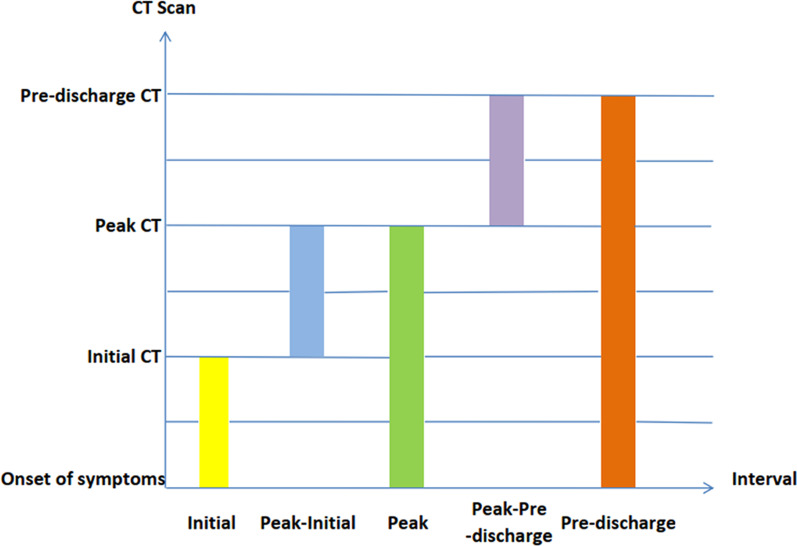
Methods: CT findings of 69 patients with COVID-19 were evaluated on initial CT, peak CT, and pre-discharge CT. CT pattern were divided into four types on CT progression. Lesion percentage of pulmonary lobe (lobe score) was graded. Correlation analysis was made between scores and intervals. 53 patients were followed up by CT.
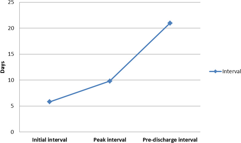
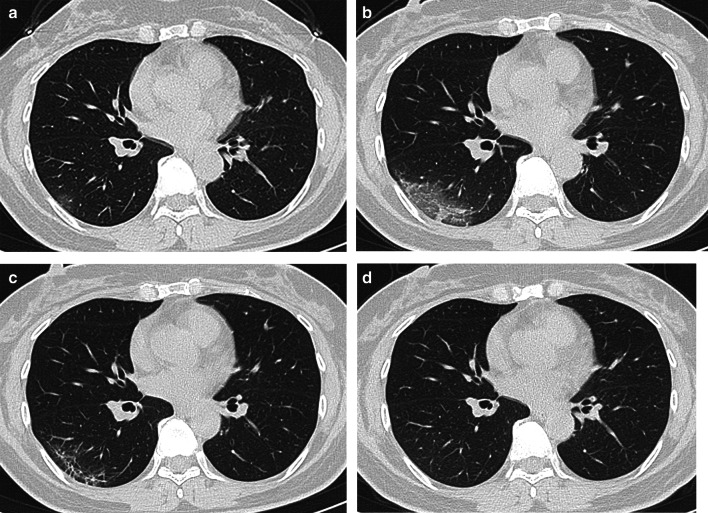
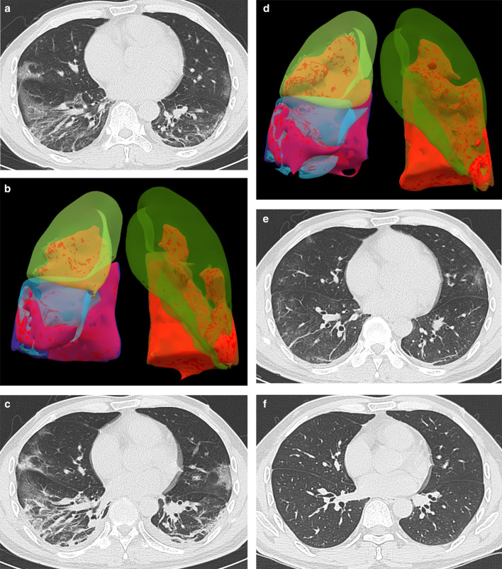
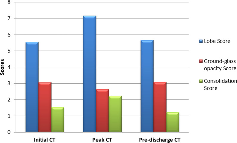
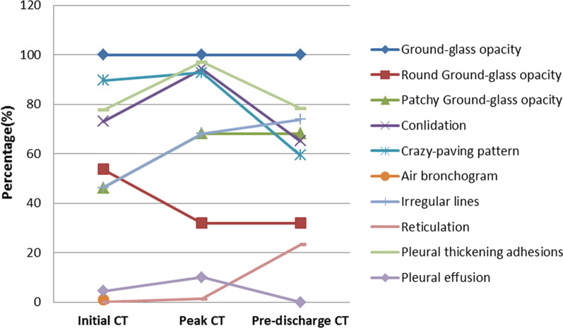
Results: Among 69 patients, 33.3% exhibited improvement pattern, 65.2% peak pattern, 1.5% deterioration pattern, and 0% fluctuation pattern. The lobe scores were positively correlated with most of intervals. It was more common to observe consolidation, pleural thickening and pleural effusion on the peak CT, and irregular line and reticulation on pre-discharge CT. The peak-initial interval were shortened when the initial CT with consolidation and pleural thickening. The intervals were extended when the irregular lines appeared on peak CT and reticulation on pre-discharge CT. Among 53 follow-up patients, 37.7% showed normal chest CT, and 62.3% showed viral pneumonia remained that mainly included GGO (100.0%) and irregular lines (33.3%).
Conclusions: COVID-19 displayed different appearances on CT as progressing. The peak pattern was the most common progression pattern. The CT appearances showed closely related to the intervals. The COVID-19 pneumonia can be remained or completely absorbed on CT with follow-up.
Keywords: COVID-19; Infectious disease medicine; Multidetector computed tomography; Pneumonia.
Conflict of interest statement
All financial and non-financial competing interests must be declared in this section.
Figures
Similar articles
-
CT findings of COVID-19 in follow-up: comparison between progression and recovery.Diagn Interv Radiol. 2020 Jul;26(4):301-307. doi: 10.5152/dir.2019.20176. Diagn Interv Radiol. 2020. PMID: 32436847 Free PMC article.
-
Prediction of the Development of Pulmonary Fibrosis Using Serial Thin-Section CT and Clinical Features in Patients Discharged after Treatment for COVID-19 Pneumonia.Korean J Radiol. 2020 Jun;21(6):746-755. doi: 10.3348/kjr.2020.0215. Korean J Radiol. 2020. PMID: 32410413 Free PMC article.
-
[Spatial and temporal distribution and predictive value of chest CT scoring in patients with COVID-19].Zhonghua Jie He He Hu Xi Za Zhi. 2021 Mar 12;44(3):230-236. doi: 10.3760/cma.j.cn112147-20200522-00626. Zhonghua Jie He He Hu Xi Za Zhi. 2021. PMID: 33721937 Chinese.
-
Imaging Features of Coronavirus disease 2019 (COVID-19): Evaluation on Thin-Section CT.Acad Radiol. 2020 May;27(5):609-613. doi: 10.1016/j.acra.2020.03.002. Epub 2020 Mar 20. Acad Radiol. 2020. PMID: 32204990 Free PMC article.
-
Coronavirus Disease 2019 (COVID-19): A Systematic Review of Imaging Findings in 919 Patients.AJR Am J Roentgenol. 2020 Jul;215(1):87-93. doi: 10.2214/AJR.20.23034. Epub 2020 Mar 14. AJR Am J Roentgenol. 2020. PMID: 32174129
Cited by
-
The Additional Value of Lower Respiratory Tract Sampling in the Diagnosis of COVID-19: A Real-Life Observational Study.Diagnostics (Basel). 2022 Sep 29;12(10):2372. doi: 10.3390/diagnostics12102372. Diagnostics (Basel). 2022. PMID: 36292061 Free PMC article.
-
The Role of Ionizing Radiation for Diagnosis and Treatment against COVID-19: Evidence and Considerations.Cells. 2022 Jan 29;11(3):467. doi: 10.3390/cells11030467. Cells. 2022. PMID: 35159277 Free PMC article. Review.
-
High correlation between CT and clinical data in COVID-19 progression.J Clin Ultrasound. 2022 Mar;50(3):383-384. doi: 10.1002/jcu.23163. J Clin Ultrasound. 2022. PMID: 35277982 Free PMC article. No abstract available.
-
CT findings in "Post-Covid": residua from acute pneumonia or "Post-Covid-ILD"?Sarcoidosis Vasc Diffuse Lung Dis. 2023 Jun 29;40(2):e2023024. doi: 10.36141/svdld.v40i2.13983. Sarcoidosis Vasc Diffuse Lung Dis. 2023. PMID: 37382073 Free PMC article.
-
Follow-up computed tomography scan in post-COVID-19 pneumonia.World J Radiol. 2022 Apr 28;14(4):104-106. doi: 10.4329/wjr.v14.i4.104. World J Radiol. 2022. PMID: 35646289 Free PMC article.
References
-
- World Health Organization. https://www.who.int/emergencies/diseases/novel-coronavirus-2019/interact...! Published 31 December 2020. Accessed 30 Mar 2021.
-
- World Health Organization. https://www.who.int/zh/news-room/detail/08-04-2020-who-timeline---covid-19. Published 11 March 2020. Accessed 9 Apr 2020.
-
- World Health Organization. https://www.who.int/emergencies/diseases/novel-coronavirus-2019/situatio.... Published 30 March 2020. Accessed 9 Apr 2021.
-
- World Health Organization. https://covid19.who.int/table. Published 30 March 2021. Accessed 30 Mar 2021.
-
- National Health Commission of the Peple’s Republic of China. Consensus on guidelines for the publication of the seventh trail version of the diagnosis and treatment plan of the novel coronavirus coronavirus. http://www.nhc.gov.cn/yzygj/s7653p/202003/46c9294a7dfe4cef80dc7f5912eb19.... Published 4 March 2020. Accessed 4 Mar 2020.
Grants and funding
LinkOut - more resources
Full Text Sources
