. 2021 Jun 10;17(6):e1009574.
doi: 10.1371/journal.pgen.1009574. eCollection 2021 Jun.

Runx1 shapes the chromatin landscape via a cascade of direct and indirect targets

Affiliations

Runx1 shapes the chromatin landscape via a cascade of direct and indirect targets

Matthew R Hass et al. PLoS Genet. .

Abstract

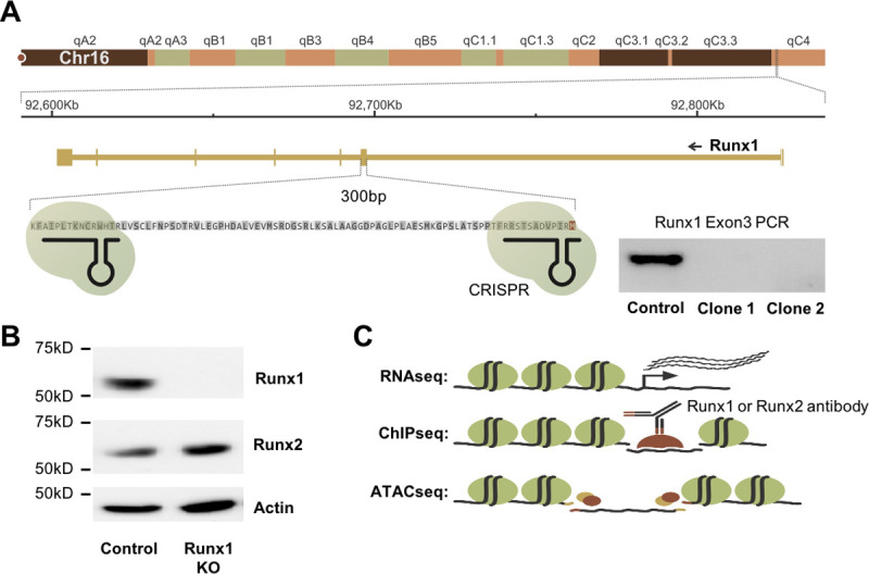
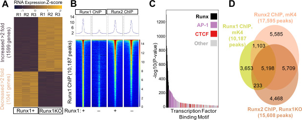
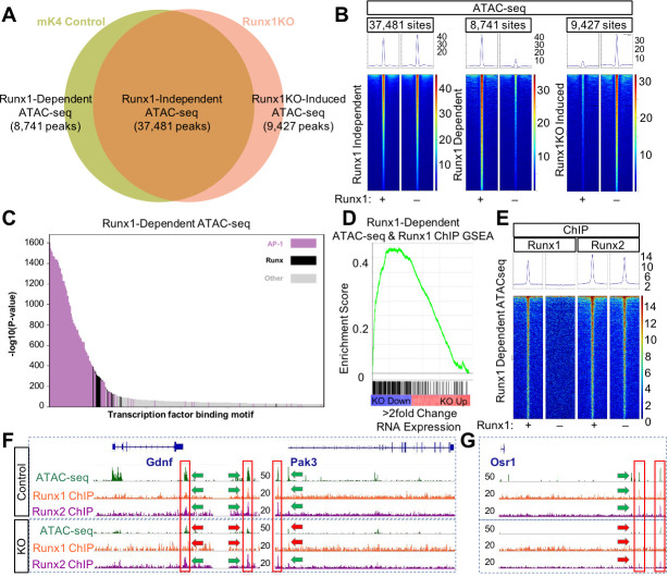
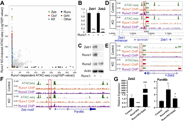
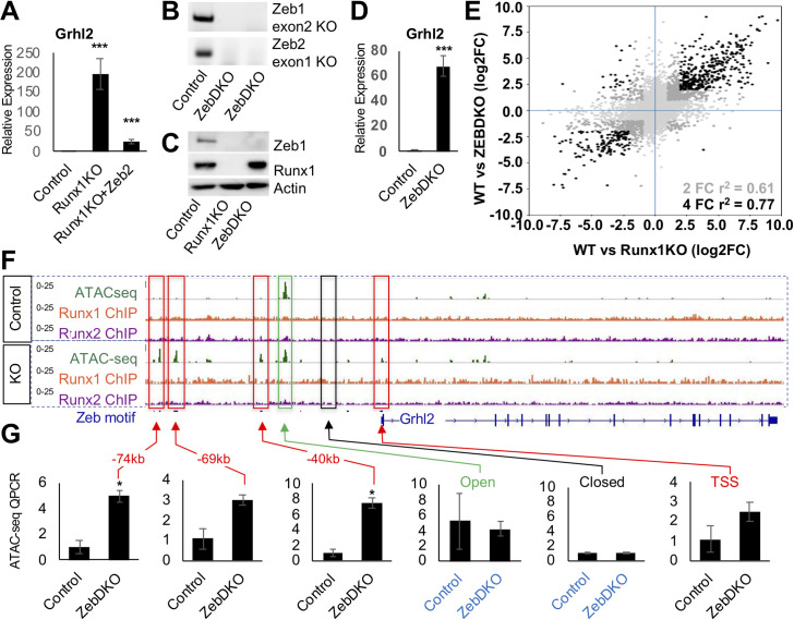
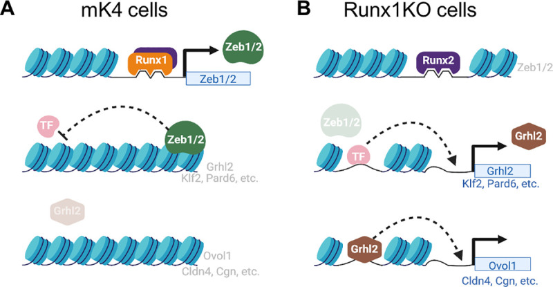
Runt-related transcription factor 1 (Runx1) can act as both an activator and a repressor. Here we show that CRISPR-mediated deletion of Runx1 in mouse metanephric mesenchyme-derived mK4 cells results in large-scale genome-wide changes to chromatin accessibility and gene expression. Open chromatin regions near down-regulated loci enriched for Runx sites in mK4 cells lose chromatin accessibility in Runx1 knockout cells, despite remaining Runx2-bound. Unexpectedly, regions near upregulated genes are depleted of Runx sites and are instead enriched for Zeb transcription factor binding sites. Re-expressing Zeb2 in Runx1 knockout cells restores suppression, and CRISPR mediated deletion of Zeb1 and Zeb2 phenocopies the gained expression and chromatin accessibility changes seen in Runx1KO due in part to subsequent activation of factors like Grhl2. These data confirm that Runx1 activity is uniquely needed to maintain open chromatin at many loci, and demonstrate that Zeb proteins are required and sufficient to maintain Runx1-dependent genome-scale repression.


Conflict of interest statement

The authors have declared that no competing interests exist.

Figures

Fig 1
Fig 1. Generation and Characterization of Runx1KO Cells.
A) Diagram of the Runx1 exon 3 region targeted for deletion using CRISPR-Cas9 and confirmation of deletion by PCR. B) Western blot showing that Runx1KO cells lack Runx1 protein but contain Runx2. C) Schematic of genomic analyses utilized to characterize Runx1KO cells.
Fig 2
Fig 2. Widespread Transcriptional Changes in Runx1KO Cells Despite Runx2 Largely Occupying the Same Regions as Runx1.
A) Heatmap of RNA-seq triplicates showing 1,705 upregulated and 1,182 downregulated genes (over 2 fold) in Runx1KO cells compared to control mK4 cells. B) Heatmaps of ChIP-seq reads mapping to Runx1 peaks from Runx1 ChIP or Runx2 ChIP in control versus Runx1KO cells with all four heatmaps being the same Runx1 ChIP peak locations ordered by the strength of the Runx1 ChIP peak. C) Graph displaying -log10 p-values of motif enrichment, revealing that Runx1 motifs are the most highly enriched motifs in the Runx1 ChIP peaks. D) Venn diagram showing the overlap of Runx1 and Runx2 ChIP peaks. Note, the lower overlap of called peaks in the Venn diagram is likely due to the Runx2 signal being just below the threshold for peak calling for many of the peaks on the lower half of the heatmap in A. RELI analysis confirmed the very significant overlap of the Runx1 and Runx2 ChIP (138.5 fold enrichment, corrected p-value 1.45 x 10−218).
Fig 3
Fig 3. Runx1 Deletion Alters Chromatin Accessibility Despite the Presence of Runx2.
A) Venn diagram of ATAC-seq peaks in control and Runx1KO cells showing the number of regions open in both cell lines (Runx1-independent), regions open only in control cells (Runx1-dependent) and regions open only in Runx1KO cells (Runx1KO-induced). B) Heatmaps of the ATAC-seq reads mapping to Runx1-independent, Runx1-dependent, and Runx1KO-induced peaks in the control and Runx1KO cells. C) Graph of the -log10 p-values of motif enrichment, displaying that Runx1-dependent ATAC-seq peaks are strongly enriched for AP-1 and Runx motifs. D) Gene set enrichment analysis showing enrichment of transcriptionally down-regulated genes (in blue and up-regulated in red) by Runx1-dependent ATAC-seq peaks that are bound by Runx1. E) Heatmap showing Runx1-Dependent ATAC-seq regions bound in Runx1 and Runx2 ChIP experiments. F) Genomic snapshots of Gdnf and Pak3 genes that are downregulated in Runx1KO cells showing open chromatin regions present in control cells but not Runx1KO cells that are bound by Runx1 and Runx2 and retain Runx2 binding in the Runx1KO cells. G) Genomic snapshot of the Runx1KO downregulated gene Osr1 that has genomic regions that lose chromatin accessibility in Runx1KO cells, which are bound by Runx1 and Runx2 in control cells, with reduced Runx2 binding in Runx1KO cells.
Fig 4
Fig 4. Runx1KO Cells Lack Zeb Repressors, Leading to the Opening of Chromatin.
A) Graph displaying p-values of transcription factor motif enrichment in Runx1-dependent versus Runx1-induced ATAC-seq, revealing that Zeb motifs are specifically enriched in Runx1KO-Induced ATAC-seq peaks. Additionally, Ctcf, Klf, and Grhl motifs are enriched in the Runx1KO-induced ATAC-seq peaks, while Runx motifs are enriched in the Runx1-dependent ATAC-seq. Note that this graph has had AP-1 motif enrichment results removed in order to focus on other motif enrichment levels (see S6A Fig for all transcription factor motifs). B) RT-qPCR showing that Runx1KO cells lose expression of Zeb1 and Zeb2. C) Western blot showing the absence of the Zeb1 protein in Runx1KO cells. D) Genomic snapshot showing a chromatin region near Zeb1 that is bound by Runx1 and Runx2 and loses chromatin accessibility in Runx1KO cells. E) Genomic snapshot of the Zeb2 locus showing a downstream potential enhancer bound by Runx1 and Runx2 that has decreased chromatin accessibility along with a loss of expression in Runx1KO cells. F) Genomic snapshot of the Zeb target gene Pard6b locus showing a promoter region containing a predicted Zeb binding site that is specifically open in Runx1KO cells. G) RT-qPCR showing that Zeb2 transient transfection of Runx1KO cells induces repression of Pard6b expression down to levels similar to those in control cells after 1 day of selection for transfected cells followed by 2 days of growth in media. (* = p-value < 0.05, ** = p < 0.005; *** = p-value < 0.0005).
Fig 5
Fig 5. Loss of Zeb Proteins Recapitulates the Opening of Chromatin and Transcriptional Induction of Runx1KO.
A) RT-qPCR confirmation of the upregulation of Grhl2 in Runx1KO cells, which is suppressed by transient transfection of Zeb2. B) Genotyping PCR showing deletions of Zeb1 exon2 and Zeb2 exon1 in ZebDKO clones. C) Western blot confirmation of loss of Zeb1 proteins in ZebDKO cells. D) RT-qPCR showing that ZebDKO cells have a dramatic increase in Grhl2 expression. E) Scatter plot showing the correlation of gene expression changes in Runx1KO and ZebDKO cells compared to control mK4 cells (light gray all genes, dark gray genes with 2 fold change in expression in both and black are genes with over 4 fold change in expression in both). F) Grhl2 loci displaying ATAC-seq regions that are specifically open in Runx1KO cells (outlined in red) that contain predicted Zeb binding sites. G) Graphs of ATAC QPCR data showing that the regions near Grhl2 that were opened in Runx1KO cells similarly become significantly more open in ZebDKO cells while control open and closed regions are similar between control and ZebDKO cells. (* = p-value < 0.05, ** = p < 0.005; *** = p-value < 0.0005).
Fig 6
Fig 6. Model of Transcription Factor Network Perturbation by Runx1-Deficiency.
A) In control mK4 cells, Runx1 induces the transcriptional repressors Zeb1 and Zeb2 that inhibit other transcriptional activators such as Grhl2, resulting in inhibition of downstream Grhl2 target genes. B) Runx1KO cells lose expression of Zeb1 and Zeb2, which derepresses their targets including Grhl2 and Klf2, which in turn leads to upregulation of their downstream targets such as Ovol1, Cldn4, and Cgn. Figure was created using BioRender.com.

Similar articles

Cited by

References

    1. Lambert SA, Jolma A, Campitelli LF, Das PK, Yin Y, Albu M, et al.. The Human Transcription Factors. Cell. 2018;175(2):598–599. doi: 10.1016/j.cell.2018.09.045 - DOI - PubMed
    1. Lee TI, Young RA. Transcriptional regulation and its misregulation in disease. Cell. 2013;152(6):1237–1251. doi: 10.1016/j.cell.2013.02.014 - DOI - PMC - PubMed
    1. Rennert J, Coffman JA, Mushegian AR, Robertson AJ. The evolution of Runx genes I. A comparative study of sequences from phylogenetically diverse model organisms. BMC Evol Biol. 2003;3:4. doi: 10.1186/1471-2148-3-4 - DOI - PMC - PubMed
    1. Mevel R, Draper JE, Lie ALM, Kouskoff V, Lacaud G. RUNX transcription factors: orchestrators of development. Development. 2019;146(17). doi: 10.1242/dev.148296 - DOI - PubMed
    1. Ito Y, Bae SC, Chuang LS. The RUNX family: developmental regulators in cancer. Nat Rev Cancer. 2015;15(2):81–95. doi: 10.1038/nrc3877 - DOI - PubMed

Publication types

MeSH terms