. 2021 Jul 1;81(13):2808-2822.e10.
doi: 10.1016/j.molcel.2021.05.018. Epub 2021 Jun 9.

Translation stress and collided ribosomes are co-activators of cGAS

Affiliations

Translation stress and collided ribosomes are co-activators of cGAS

Li Wan et al. Mol Cell. .

Abstract

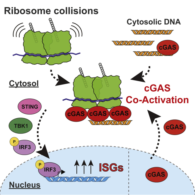
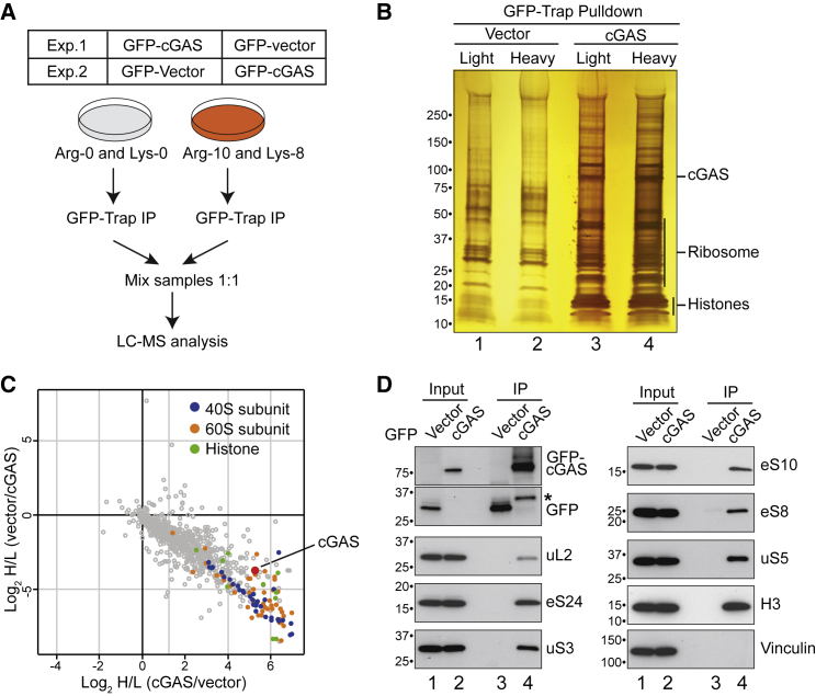
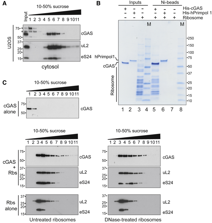
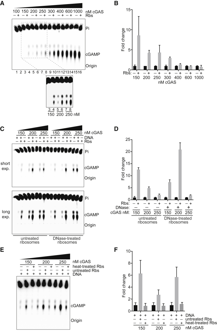
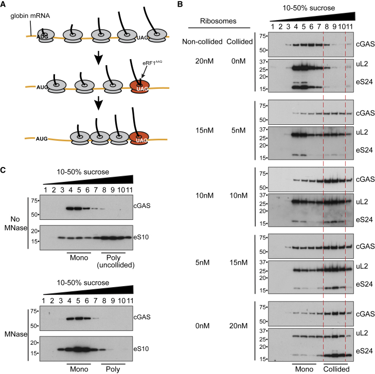
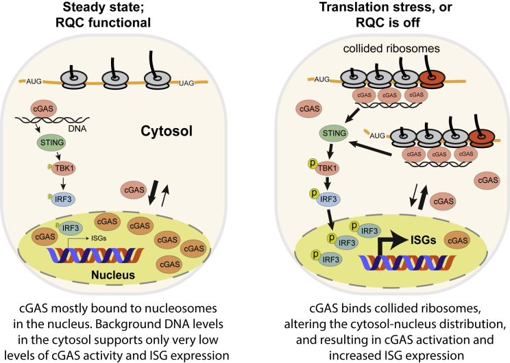
The cyclic GMP-AMP synthase-stimulator of interferon genes (cGAS-STING) pathway senses cytosolic DNA and induces interferon-stimulated genes (ISGs) to activate the innate immune system. Here, we report the unexpected discovery that cGAS also senses dysfunctional protein production. Purified ribosomes interact directly with cGAS and stimulate its DNA-dependent activity in vitro. Disruption of the ribosome-associated protein quality control (RQC) pathway, which detects and resolves ribosome collision during translation, results in cGAS-dependent ISG expression and causes re-localization of cGAS from the nucleus to the cytosol. Indeed, cGAS preferentially binds collided ribosomes in vitro, and orthogonal perturbations that result in elevated levels of collided ribosomes and RQC activation cause sub-cellular re-localization of cGAS and ribosome binding in vivo as well. Thus, translation stress potently increases DNA-dependent cGAS activation. These findings have implications for the inflammatory response to viral infection and tumorigenesis, both of which substantially reprogram cellular protein synthesis.

Keywords: ASCC3; IRF3; STING; ZNF598; cGAS; innate immunity; interferon signalling; mRNA translation; ribosome collision; ribosome-associated protein quality control.


Conflict of interest statement

Declaration of interests The authors declare no competing interests.

Figures

None
Graphical abstract
Figure 1
Figure 1
The cGAS-STING pathway is required for increased ISG expression in ASCC3-deficient cells (A) Schematic of relevant innate immunity signaling pathways. (B) Western blot analysis of IRF3 and TBK1, and their phosphorylated forms (IRF3ser396 or TBK1ser172), in cells depleted of ASCC3. (C) qRT-PCR analysis of relative ISG expression in MRC5VA cells treated with siRNAs. Error bars represent standard deviation (SD) of three technical replicates and are representative of three biological replicates. (D) Western blot analysis of IRF3p (ser396), IRF3, TBK1p (ser172), TBK1, RSAD2, IFIT2, STAT1p (tyr701), STAT1, ASCC3, STING, and MAVS in the same cells as in (C). (E) Western blot analysis of RSAD2, IFIT2, STAT1p (tyr701), STAT1, ASCC3, and cGAS in parental MRC5VA and two different CGAS knockout cell lines (KO-7 and -12) after ASCC3 knockdown. Asterisks denote non-specific bands. (F) As in (E), but in U2OS cells. (G) qRT-PCR analysis of relative ISG expression in U2OS cells transfected with the indicated siRNAs. Error bars represent SD of three technical replicates and are representative of three biological replicates. (H) As in (G), but also using U2OS CGAS KO-16 cells. See also Figures S1–S4.
Figure 2
Figure 2
The cGAS interactome (A) Strategy for SILAC-based quantitative mass spectrometry. (B) Silver staining showing GFP-associated factors (Vector [control]) and GFP-cGAS-associated factors (cGAS), respectively. (C) Scatterplots of Log2 SILAC ratios for the cGAS interactome. Small ribosomal proteins are marked in blue, large ribosomal proteins in orange, and histone proteins in green. (D) Validation by immunoprecipitation (IP)-western blotting with the indicated antibodies. The two upper panels on the left are from same the anti-GFP blot. Asterisk denotes a likely GFP-cGAS degradation product.
Figure 3
Figure 3
Evidence for a direct cGAS-ribosome interaction (A) Cytosol from U2OS cells was separated by sucrose gradient sedimentation, and fractions were immunoblotted for cGAS and representative ribosome subunits (ul2 and eS24). Asterisks denote non-specific bands. (B) Purified ribosomes were incubated with Ni-NTA agarose, Ni-NTA agarose with immobilized human recombinant cGAS-8his, or hPrimpol1-8his (control). After washing, the eluate was analyzed by SDS-PAGE and Coomassie staining. (C) Western blot analysis of cGAS, ribosomes (both untreated and DNase-treated ribosomes), or cGAS-ribosome complex, separated by sucrose gradient sedimentation. See also Figure S5.
Figure 4
Figure 4
Ribosomes stimulate DNA-dependent cGAS activity in vitro (A) Autoradiograph of cGAS-mediated cGAMP synthesis in the presence of different concentrations of cGAS with or without ribosomes; all are in the presence of 1 μg herring testis DNA, which is saturating (see Figures S6A and S6B). (B) Quantification of data in (A) by Fiji. Error bars indicate SD of duplicate replicates. (C) As in (A), but with untreated ribosomes or DNase-treated ribosomes. (D) Quantification of the data from (C), as in (B). (E) As in (A), but with untreated or heat-treated ribosomes. (F) Quantification of the data from (E), as in (B). See also Figures S6 and S7.
Figure 5
Figure 5
cGAS preferentially interacts with collided ribosomes (A) Strategy to generate collided ribosomes using an in vitro translation reaction in rabbit reticulocyte lysate (Juszkiewicz et al., 2018). (B) cGAS was incubated with a mixture of collided and non-collided ribosomes at different ratios, separated by 10%–50% sucrose gradient fractionation, and then analyzed by western blotting. Collided ribosomes (fractions 8, 9, and 10) are indicated by dashed lines. (C) Western blot analysis of MRC5VA cytosol fractionated by sucrose gradient sedimentation, with or without prior incubation with micrococcal nuclease (MNase) to digest polysomes (poly) to monosomes (mono).
Figure 6
Figure 6
Conditions that result in collided ribosomes induce cytosolic localization of cGAS (A) U2OS cells stably expressing GFP-cGAS were transfected with the indicated siRNA. Cells were fixed, stained with eS8 antibody or with DAPI, and imaged by confocal fluorescence microscopy. Scale bar: 10 μm. (B) As in (A), but after treatment with the indicated drug regimens. (C) As in (A) and (B), but after acute heat shock treatment. Quantification of Figures S8A–S8C. (D) Analysis of the interaction between cGAS and ribosomes using the in situ proximity ligation assay (PLA) before and after heat shock in U2OS cGAS KO cells stably expressing FLAG-hemagglutinin (HA)-tagged GAS. Cells were fixed, incubated with the indicated antibodies, and visualized according to instruction of Dulink In Situ Kit. The PLA signal was detected by confocal fluorescence microscopy. Scale bar: 10 μm. (E) Quantitative analysis of (D). Two-tailed t test, ∗∗∗∗p < 0.0001. Error bars represent SD of puncta per cell from 80 cells per condition. (F) qRT-PCR analysis of relative ISG expression in U2OS cells treated with heat shock. Error bars represent SD of three technical replicates and are representative of three biological replicates. (G) As in (F), but cells are also treated with translation inhibitor cycloheximide (CHX). ns, no significant. See also Figure S8 for quantification.
Figure 7
Figure 7
Working model for the cGAS response to translation stress (A) In steady state or when the RQC is functional, cGAS predominantly binds to nucleosomes in the nucleus. Background DNA levels in the cytosol support very low levels of cGAS activity and background ISG expression. (B) During translation stress or when the RQC is off, cGAS binds to collided ribosomes, which alters the cytosol-nucleus distribution and results in cGAS activation and increased ISG expression.

References

    1. Ablasser A., Chen Z.J. cGAS in action: Expanding roles in immunity and inflammation. Science. 2019;363:eaat8657. - PubMed
    1. Ablasser A., Goldeck M., Cavlar T., Deimling T., Witte G., Röhl I., Hopfner K.P., Ludwig J., Hornung V. cGAS produces a 2′-5′-linked cyclic dinucleotide second messenger that activates STING. Nature. 2013;498:380–384. - PMC - PubMed
    1. Ablasser A., Hertrich C., Waßermann R., Hornung V. Nucleic acid driven sterile inflammation. Clin. Immunol. 2013;147:207–215. - PubMed
    1. Andreeva L., Hiller B., Kostrewa D., Lässig C., de Oliveira Mann C.C., Jan Drexler D., Maiser A., Gaidt M., Leonhardt H., Hornung V., Hopfner K.P. cGAS senses long and HMGB/TFAM-bound U-turn DNA by forming protein-DNA ladders. Nature. 2017;549:394–398. - PubMed
    1. Arquint C., Nigg E.A. STIL microcephaly mutations interfere with APC/C-mediated degradation and cause centriole amplification. Curr. Biol. 2014;24:351–360. - PubMed

Publication types

Substances