. 2021 Aug;148(2):368-380.e3.
doi: 10.1016/j.jaci.2021.05.032. Epub 2021 Jun 7.

Chemokines and eicosanoids fuel the hyperinflammation within the lungs of patients with severe COVID-19

Affiliations

Chemokines and eicosanoids fuel the hyperinflammation within the lungs of patients with severe COVID-19

Younes Zaid et al. J Allergy Clin Immunol. 2021 Aug.

Abstract

Background: Severe acute respiratory syndrome coronavirus 2 (SARS-CoV-2) infection can lead to a variety of clinical outcomes, ranging from the absence of symptoms to severe acute respiratory disease and ultimately death. A feature of patients with severe coronavirus disease 2019 (COVID-19) is the abundance of inflammatory cytokines in the blood. Elevated levels of cytokines are predictive of infection severity and clinical outcome. In contrast, studies aimed at defining the driving forces behind the inflammation in lungs of subjects with severe COVID-19 remain scarce.

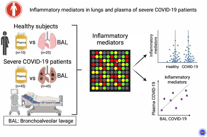
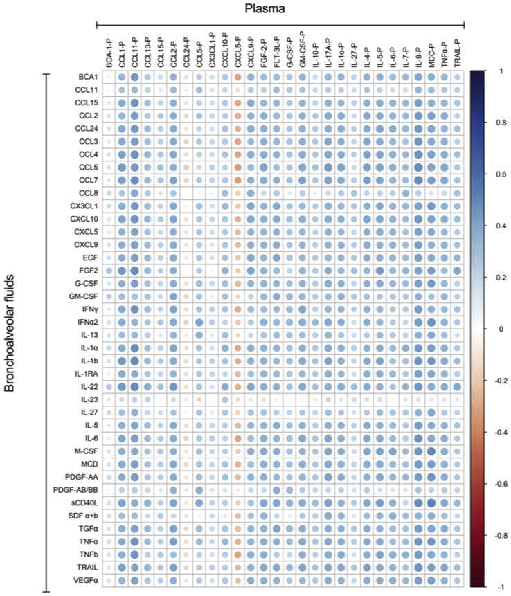
Objective: Our aim was to analyze and compare the plasma and bronchoalveolar lavage (BAL) fluids of patients with severe COVID-19 (n = 45) for the presence of cytokines and lipid mediators of inflammation (LMIs).

Methods: Cytokines were measured by using Luminex multiplex assay, and LMIs were measured by using liquid chromatography-tandem mass spectrometry.

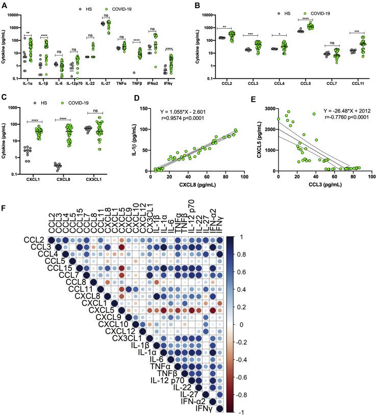
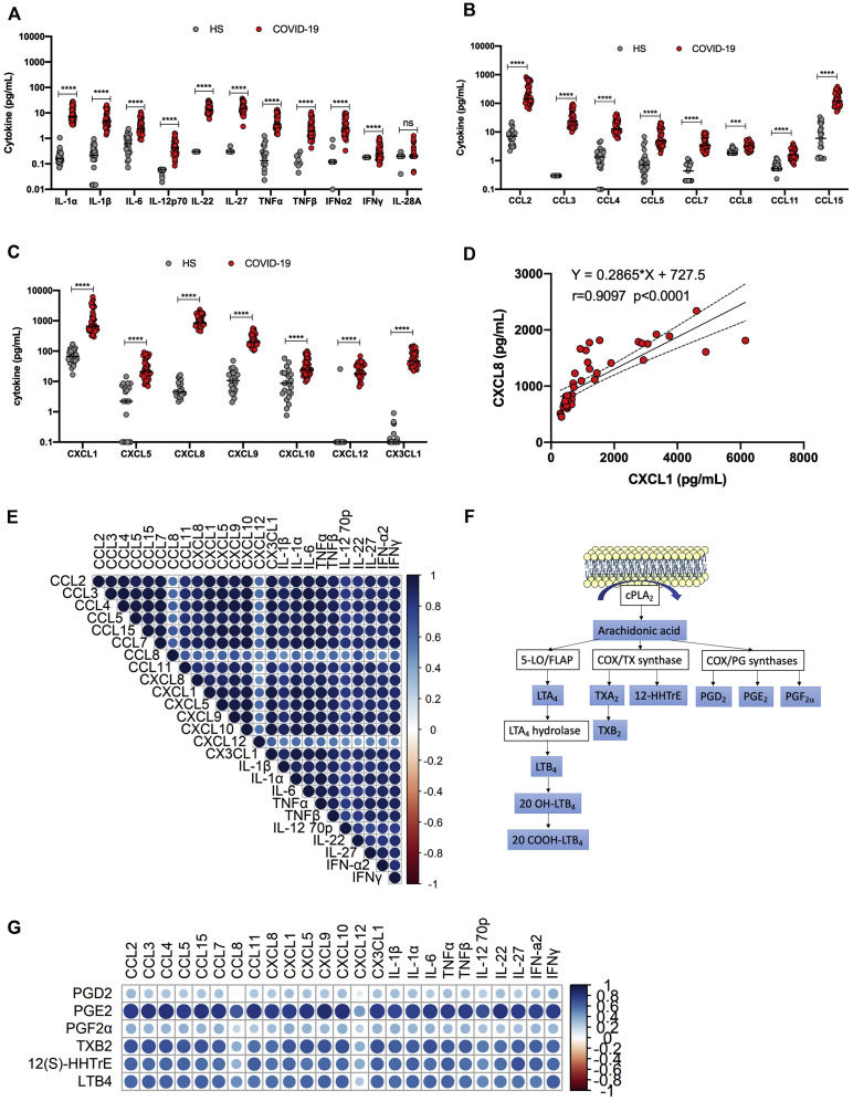
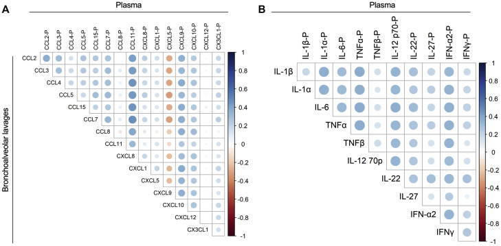
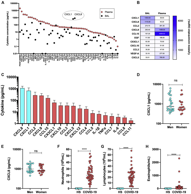
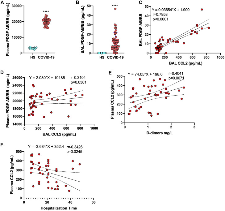
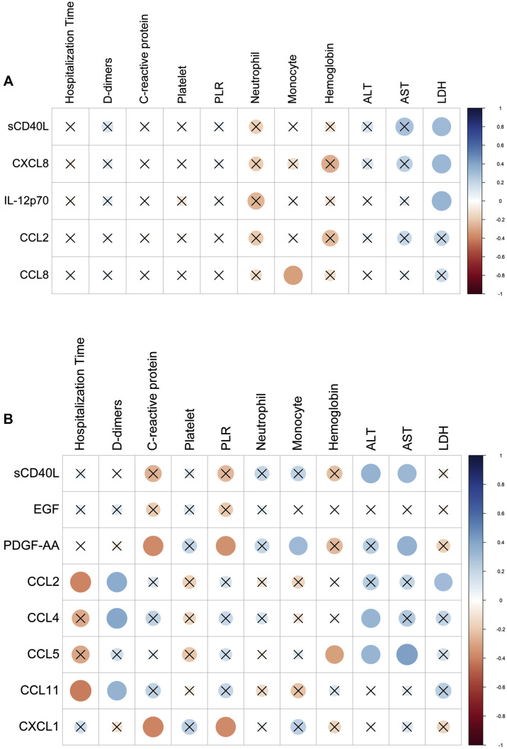
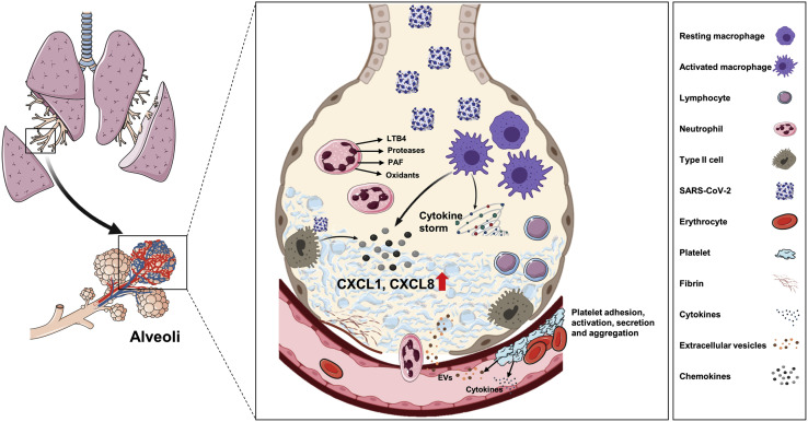
Results: We revealed high concentrations of numerous cytokines, chemokines, and LMIs in the BAL fluid of patients with severe COVID-19. Of the 13 most abundant mediators in BAL fluid, 11 were chemokines, with CXCL1 and CXCL8 being 200 times more abundant than IL-6 and TNF-α. Eicosanoid levels were also elevated in the lungs of subjects with severe COVID-19. Consistent with the presence chemotactic molecules, BAL fluid samples were enriched for neutrophils, lymphocytes, and eosinophils. Inflammatory cytokines and LMIs in plasma showed limited correlations with those present in BAL fluid, arguing that circulating inflammatory molecules may not be a reliable proxy of the inflammation occurring in the lungs of patients with severe COVID-19.

Conclusions: Our findings indicate that hyperinflammation of the lungs of patients with severe COVID-19 is fueled by excessive production of chemokines and eicosanoids. Therapeutic strategies to dampen inflammation in patients with COVID-19 should be tailored accordingly.

Keywords: ARDS; COVID-19; SARS-CoV-2; chemokines; eicosanoids; inflammatory cytokines; lipid mediators of inflammation.


Figures

None
Graphical abstract
Fig 1
Fig 1
Analysis of cytokines, chemokines, and growth factors in plasma of patients with severe COVID-19 (n = 45) and healthy subjects (n = 10). A-C, Plasmatic concentrations of various cytokines (A), CC chemokines (B), and CXC chemokines (C) in controls and subjects with severe COVID-19. D, Example of positive correlation between IL-1β and CXCL8. E, Example of negative correlation between CXCL5 and CCL3. F, Summary of Spearman correlations between plasma analytes of subjects with severe COVID-19. Results are presented as scattered plots with median concentrations shown by horizontal bars. In this figure, COVID-19 refers to patients with severe COVID-19. ∗∗P < .005 and ∗∗∗∗P < .0001, as determined by using the Mann-Whitney nonparametric test. HS, Healthy subject.
Fig 2
Fig 2
Analysis of cytokines, chemokines, and growth factors in BAL fluid of patients with severe COVID-19 (n = 45) and healthy subjects (n = 25). A, BAL fluid concentrations of various cytokines and interferons. B, BAL fluid concentrations of various CC chemokines. C, BAL fluid concentrations of various CXC chemokines. D, Example of positive correlation between CXCL8 and CXCL1. E, Summary of Spearman correlations between cytokines in BAL fluid of subjects with severe COVID-19. F, Diagram showing generation of arachidonic acid following membrane phospholipid hydrolysis by cPLA2 and subsequent processing into various LMs. G, Summary of Spearman correlations between LMs and cytokines in BAL fluid of subjects with severe COVID-19. In this figure, COVID-19 refers to patients with severe COVID-19. Results are presented as scattered plots with median concentrations shown by horizontal bars. ∗∗∗∗P < .0001, as determined using the Mann-Whitney nonparametric test. HS, Healthy subject.
Fig 3
Fig 3
Spearman correlations between cytokines in BAL fluid and plasma of subjects with severe COVID-19. A, CC and CXC chemokines correlations. B, Cytokines correlations. Blue indicates positive correlations, and red indicates negative correlation.
Fig 4
Fig 4
Comparison of cytokine levels in BAL fluid and plasma. A, The absolute median concentrations of cytokines, chemokines, and growth factors in BAL fluid and plasma. B, Heat map representation of cytokine, chemokine, and growth factor levels in BAL fluid and plasma of patients with severe COVID-19. Numbers in box represent the mean levels in detected. C, The absolute concentration of various chemokines and cytokines in BAL fluid of patients with severe COVID-19. D, CXCL1 levels in BAL fluid of male and female subjects with subjects with severe COVID-19. E CXCL8 levels in BAL fluid of male and female subjects with severe COVID-19. F-H, Neutrophil (F), lymphocyte (G), and eosinophil (H) contents of BAL fluid of healthy subjects and patients with severe COVID-19. C-E, Results are expressed as medians ± SDs. F-H, Results are expressed as mean cell counts ± SDs. ∗∗∗∗P < .0001, as determined using the Mann-Whitney nonparametric test.
Fig 5
Fig 5
PDGF-AB/BB and CCL2 in BAL fluid and plasma. A, Concentrations of PDGF-AB/BB in plasma of healthy individuals and patients with severe COVID-19. B, Concentrations of PDGF-AB/BB in BAL fluid of healthy individuals and patients with severe COVID-19. C, Correlation between PDBF-AB/BB and CCL2 in BAL fluid of patients with severe COVID-19. D, Correlation between PDBF-AB/BB in plasma and CCL2 in BAL fluid of patients with severe COVID-19. E, Correlation between CCL2 and d-dimer levels in plasma of patients with severe COVID-19. F, Correlation between CCL2 and duration of hospitalization of patients with severe COVID-19. Nonparametric Spearman correlation coefficients were calculated to study the association between cytokines. ∗∗∗∗P < .0001, as determined by using the Mann-Whitney nonparametric test.
Fig 6
Fig 6
Spearman correlation of levels of cytokines, chemokines, and LMs with biochemical and hematologic parameters and duration of hospitalization of patients with severe COVID-19. A, Spearman correlation of levels of cytokines and chemokines in BAL fluid with biochemical and hematologic parameters and duration of hospitalization of patients with severe COVID-19. B, Spearman correlation of levels of cytokines and chemokines in plasma with biochemical and hematologic parameters and duration of hospitalization of patients with patients with severe COVID-19. X denotes nonsignificant correlations.
Fig 7
Fig 7
Proposed lung pathophysiology associated with SARS-CoV-2 infection. The SARS-CoV-2 virus infects type II pneumocytes and activates macrophages, causing the release of several cytokines and chemokines, including CXCL8 and CXCL1. The viral infection itself and various cytokines cause endothelial activation and dysfunction, resulting in vascular inflammation and permeability. Immune cells such as neutrophils and lymphocytes migrate into alveoli in response to the secreted chemoattractants and enhance lung inflammation. Damaged endothelial cell barrier causes platelet activation and aggregation as well as microthrombi formation. Together, inflammation, edema, and microthrombi cause ARDS.
Fig E1
Fig E1
Spearman correlation between cytokines and chemokines in plasma and in BAL fluids of subjects with severe COVID-19.

References

    1. Wu F., Zhao S., Yu B., Chen Y.M., Wang W., Song Z.G., et al. A new coronavirus associated with human respiratory disease in China. Nature. 2020;579:265–269. - PMC - PubMed
    1. Qin C., Zhou L., Hu Z., Zhang S., Yang S., Tao Y., et al. Dysregulation of immune response in patients with coronavirus 2019 (COVID-19) in Wuhan, China. Clin Infect Dis. 2020;71:762–768. - PMC - PubMed
    1. Wu C., Chen X., Cai Y., Xia J., Zhou X., Xu S., et al. Risk factors associated with acute respiratory distress syndrome and death in patients with Coronavirus disease 2019 pneumonia in Wuhan, China. JAMA Intern Med. 2020;180:934–943. - PMC - PubMed
    1. Hoffmann M., Kleine-Weber H., Schroeder S., Kruger N., Herrler T., Erichsen S., et al. SARS-CoV-2 cell entry depends on ACE2 and TMPRSS2 and is blocked by a clinically proven protease inhibitor. Cell. 2020;181:271–280.e8. - PMC - PubMed
    1. Chen N., Zhou M., Dong X., Qu J., Gong F., Han Y., et al. Epidemiological and clinical characteristics of 99 cases of 2019 novel coronavirus pneumonia in Wuhan, China: a descriptive study. Lancet. 2020;395:507–513. - PMC - PubMed

MeSH terms