Whole-body magnetic resonance imaging (WB-MRI) with diffusion-weighted whole-body imaging with background body signal suppression (DWIBS) in prostate cancer: Prevalence and clinical significance of incidental findings
- PMID: 34111963
- PMCID: PMC8978253
- DOI: 10.1259/bjr.20210459
Whole-body magnetic resonance imaging (WB-MRI) with diffusion-weighted whole-body imaging with background body signal suppression (DWIBS) in prostate cancer: Prevalence and clinical significance of incidental findings
Abstract
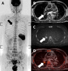
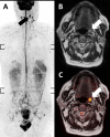
Objective: Diffusion-weighted whole-body imaging with background body signal suppression (DWIBS) is now recommended as a first-line staging modality in prostate cancer patients, and the widespread use of DWIBS may lead to an increased frequency of incidental findings. The aim of this study was to evaluate the prevalence and clinical significance of incidental findings on whole-body magnetic resonance imaging (WB-MRI) with DWIBS.
Methods: Data from 124 patients with prostate cancer (age, 76.5 ± 5.6 years), who underwent 1.5 T WB-MRI with STIR, TSE-T2, TSE-T1, In/Out GRE, and DWIBS sequences, were retrospectively analyzed. Findings unrelated to prostate cancer were considered as incidental findings and categorized into two groups based on their clinical implications as follows: imaging follow-up or additional examinations was required (significant incidental findings) and no need to additional work-up (non-significant incidental findings). A chi-square test was performed to compare the differences in the prevalence of significant incidental findings based on age (≤75 and>75 years old).
Results: A total of 334 incidental findings were found with 8.1% (n = 27) as significant incidental findings. Significant incidental findings were more frequent in patients over 75 years old than those of 75 years old or younger (28.6% vs 11.1%, p = 0.018).
Conclusion: Clinically significant incidental findings, which required imaging follow-up or additional examinations, were commonly observed in prostate cancer patients on WB-MRI/DWIBS.
Advances in knowledge: Some incidental findings were clinically significant that may lead to changes in treatment strategy. Checking the entire organ carefully for abnormalities and reporting any incidental findings detected are important.
Figures


References
-
- Takahara T, Imai Y, Yamashita T, Yasuda S, Nasu S, Van Cauteren M. Diffusion weighted whole body imaging with background body signal suppression (DWIBS): technical improvement using free breathing, stir and high resolution 3D display. Radiat Med 2004; 22: 275–82. - PubMed
-
- Takenaka D, Ohno Y, Matsumoto K, Aoyama N, Onishi Y, Koyama H, et al. . Detection of bone metastases in non-small cell lung cancer patients: comparison of whole-body diffusion-weighted imaging (DWI), whole-body MR imaging without and with DWI, whole-body FDG-PET/CT, and bone scintigraphy. J Magn Reson Imaging 2009; 30: 298–308. doi: 10.1002/jmri.21858 - DOI - PubMed
-
- Laurent V, Trausch G, Bruot O, Olivier P, Felblinger J, Régent D. Comparative study of two whole-body imaging techniques in the case of melanoma metastases: advantages of multi-contrast MRI examination including a diffusion-weighted sequence in comparison with PET-CT. Eur J Radiol 2010; 75: 376–83. doi: 10.1016/j.ejrad.2009.04.059 - DOI - PubMed
MeSH terms
LinkOut - more resources
Full Text Sources
Medical

