

Transcriptomic analysis of shell repair and biomineralization in the blue mussel, Mytilus edulis
- PMID: 34112105
- PMCID: PMC8194122
- DOI: 10.1186/s12864-021-07751-7
Transcriptomic analysis of shell repair and biomineralization in the blue mussel, Mytilus edulis
Abstract
Background: Biomineralization by molluscs involves regulated deposition of calcium carbonate crystals within a protein framework to produce complex biocomposite structures. Effective biomineralization is a key trait for aquaculture, and animal resilience under future climate change. While many enzymes and structural proteins have been identified from the shell and in mantle tissue, understanding biomieralization is impeded by a lack of fundamental knowledge of the genes and pathways involved. In adult bivalves, shells are secreted by the mantle tissue during growth, maintenance and repair, with the repair process, in particular, amenable to experimental dissection at the transcriptomic level in individual animals.
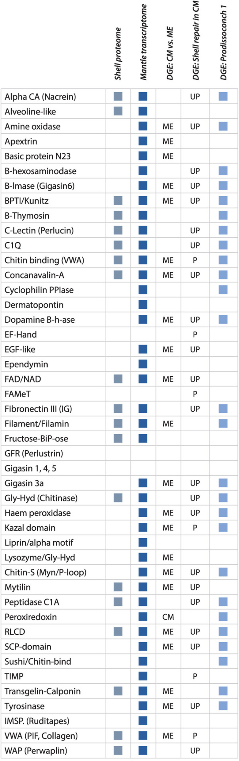
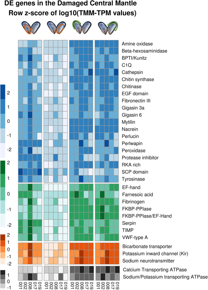
Results: Gene expression dynamics were explored in the adult blue mussel, Mytilus edulis, during experimentally induced shell repair, using the two valves of each animal as a matched treatment-control pair. Gene expression was assessed using high-resolution RNA-Seq against a de novo assembled database of functionally annotated transcripts. A large number of differentially expressed transcripts were identified in the repair process. Analysis focused on genes encoding proteins and domains identified in shell biology, using a new database of proteins and domains previously implicated in biomineralization in mussels and other molluscs. The genes implicated in repair included many otherwise novel transcripts that encoded proteins with domains found in other shell matrix proteins, as well as genes previously associated with primary shell formation in larvae. Genes with roles in intracellular signalling and maintenance of membrane resting potential were among the loci implicated in the repair process. While haemocytes have been proposed to be actively involved in repair, no evidence was found for this in the M. edulis data.
Conclusions: The shell repair experimental model and a newly developed shell protein domain database efficiently identified transcripts involved in M. edulis shell production. In particular, the matched pair analysis allowed factoring out of much of the inherent high level of variability between individual mussels. This snapshot of the damage repair process identified a large number of genes putatively involved in biomineralization from initial signalling, through calcium mobilization to shell construction, providing many novel transcripts for future in-depth functional analyses.
Keywords: Bivalve; Calcium; Haemocytes; Mollusc; Shell matrix proteins.
Conflict of interest statement
The authors declare that they have no competing interests.
Figures


Similar articles
-
A Bivalve Biomineralization Toolbox.Mol Biol Evol. 2021 Aug 23;38(9):4043-4055. doi: 10.1093/molbev/msab153. Mol Biol Evol. 2021. PMID: 34014311 Free PMC article.
-
A shell regeneration assay to identify biomineralization candidate genes in mytilid mussels.Mar Genomics. 2016 Jun;27:57-67. doi: 10.1016/j.margen.2016.03.011. Epub 2016 Apr 12. Mar Genomics. 2016. PMID: 27083865
-
Characterization of the mantle transcriptome in bivalves: Pecten maximus, Mytilus edulis and Crassostrea gigas.Mar Genomics. 2016 Jun;27:9-15. doi: 10.1016/j.margen.2016.04.003. Epub 2016 May 7. Mar Genomics. 2016. PMID: 27160853
-
Deciphering mollusc shell production: the roles of genetic mechanisms through to ecology, aquaculture and biomimetics.Biol Rev Camb Philos Soc. 2020 Dec;95(6):1812-1837. doi: 10.1111/brv.12640. Epub 2020 Jul 31. Biol Rev Camb Philos Soc. 2020. PMID: 32737956 Review.
-
Sea shell diversity and rapidly evolving secretomes: insights into the evolution of biomineralization.Front Zool. 2016 Jun 7;13:23. doi: 10.1186/s12983-016-0155-z. eCollection 2016. Front Zool. 2016. PMID: 27279892 Free PMC article. Review.
Cited by
-
De novo genome assembly and genome skims reveal LTRs dominate the genome of a limestone endemic Mountainsnail (Oreohelix idahoensis).BMC Genomics. 2022 Dec 2;23(1):796. doi: 10.1186/s12864-022-09000-x. BMC Genomics. 2022. PMID: 36460988 Free PMC article.
-
In the footsteps of sea stars: deciphering the catalogue of proteins involved in underwater temporary adhesion.Open Biol. 2022 Aug;12(8):220103. doi: 10.1098/rsob.220103. Epub 2022 Aug 17. Open Biol. 2022. PMID: 35975651 Free PMC article.
-
Metabolic profiling of Mytilus coruscus mantle in response of shell repairing under acute acidification.PLoS One. 2023 Oct 27;18(10):e0293565. doi: 10.1371/journal.pone.0293565. eCollection 2023. PLoS One. 2023. PMID: 37889901 Free PMC article.
-
The Mantle Transcriptome of Chamelea gallina (Mollusca: Bivalvia) and Shell Biomineralization.Animals (Basel). 2022 May 6;12(9):1196. doi: 10.3390/ani12091196. Animals (Basel). 2022. PMID: 35565623 Free PMC article.
-
Transcriptomic response of Mytilus coruscus mantle to acute sea water acidification and shell damage.Front Physiol. 2023 Oct 26;14:1289655. doi: 10.3389/fphys.2023.1289655. eCollection 2023. Front Physiol. 2023. PMID: 37954445 Free PMC article.
References
-
- Checa AG. Physical and biological determinants of the fabrication of molluscan shell microstructures. Front Mar Sci. 2018;5:353. doi: 10.3389/fmars.2018.00353. - DOI
-
- Yarra T: Transcriptional Profiling of Shell Calcification in Bivalves. PhD thesis, University of Edinburgh, UK, 2018.
MeSH terms
Grants and funding
LinkOut - more resources
Full Text Sources
