Case Reports
. 2021 Jun 10;17(1):215.
doi: 10.1186/s12917-021-02921-x.

Acute fasciolosis in an alpaca: a case report

Affiliations
Case Reports

Acute fasciolosis in an alpaca: a case report

C J Hayes et al. BMC Vet Res. .

Abstract

Background: The popularity of new world camelids, particularly alpacas, is growing rapidly in Ireland, presenting a clinical challenge to veterinary practitioners who may not have worked with these species previously. To the authors' knowledge, the clinical course of a case of acute fasciolosis in an alpaca has not previously been reported, and fasciolosis has not been reported at all in alpacas in Ireland, making this case report a valuable addition to the current literature.

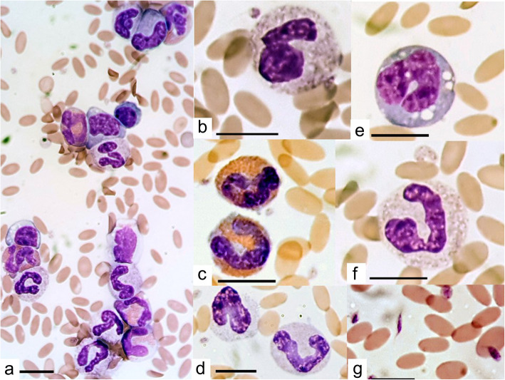
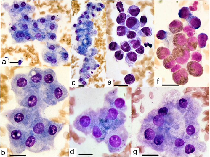
Case presentation: A three-year-old male castrated huacaya alpaca was admitted to UCD Veterinary Hospital with a two-day history of colic and tenesmus. He had been treated with albendazole, dexamethasone and potentiated amoxycillin by the referring veterinary practitioner with no response. On initial clinical exam, sensitivity to abdominal palpation was the only abnormality. However, the alpaca proceeded to show abnormal lying positions, tenesmus and reduced faecal output over the next 24 h. A general blood panel demonstrated moderate anaemia, marked hyperglobulinaemia and moderately increased hepatocellular and hepatobiliary enzyme activity. Abdominal radiography revealed enlargement of the first forestomach compartment without evidence of gastrointestinal obstruction or peritonitis. An abdominal ultrasound exam revealed an elongated, heterogenous mass in the caudoventral abdomen that appeared to be contiguous with the liver. FNA of this mass revealed that it was in fact a liver lobe with biliary stasis and inflammation. Faecal sedimentation demonstrated Fasciola hepatica eggs. In spite of treatment with triclabendazole and supportive treatment including blood transfusion, the alpaca's condition continued to deteriorate and he was euthanised. On post-mortem exam, acute fasciolosis was diagnosed.

Conclusions: The clinical presentation and course of a case of acute fasciolosis in an individual alpaca is described, including the results of a range of diagnostic tests that were carried out. The final diagnosis is supported by a description of post-mortem findings. This information will serve as a resource for veterinary practitioners involved in the diagnosis and treatment of similar cases.

Keywords: Acute fasciolosis; Alpaca; Liver fluke; New World camelid.


Conflict of interest statement

The authors declare that they have no competing interests.

Figures

Fig. 1
Fig. 1
Blood smear taken on day five of hospitalisation of an alpaca diagnosed with acute fasciolosis. Photomicrograph (a) demonstrates decreased erythrocyte density, predominance of eosinophils and immature neutrophils. Other photomicrographs demonstrate close-ups of toxic neutrophils (b,f), eosinophils (c), immature neutrophils (d), monocytes (e) and pleomorphic platelets (g). Images taken using Motic BA410 microscope, Motic Moticam 10 (10.0 mp) camera and Motic Images Pluse 2.0 image processing software, all purchased from Motic Europe, Barcelona, Spain. 100 erythrocytes were measured using the Motic calibration cytometer and their mean size was found to be 6.23 μm with a standard deviation of 0.56 μm and range 4.7 to 7.7 μm. Erythrocyte size was normally distributed. A scale bar in the bottom left of each photomicrograph indicates 10 μm
Fig. 2
Fig. 2
Lateral abdominal radiograph of a three-year old male huacaya alpaca diagnosed with acute fasciolosis. Sub-figure a shows the dorsal thorax and abdomen. Sub-figure b shows the caudodorsal abdomen. Sub-figure c shows the caudoventral abdomen. Sub-figure d shows the cranioventral thorax and abdomen
Fig. 3
Fig. 3
Ultrasound image of a caudoventral abdominal mass identified in an alpaca diagnosed with acute fasciolosis
Fig. 4
Fig. 4
Cytological findings from ultrasound-guided, fine-needle aspiration of an abdominal mass in an alpaca. Photomicrographs a to d and g demonstrate monomorphic hepatocytes with mild vacuolar change (a, c) cell swelling (a, g), mild accumulation of tiny, blue-green, granules conistent with bile (a, c, d) and binucleation (a). Photomicrographs E and F show inflammation with increased numbers of neutrophils and mononuclear cells (e) and eosinophilis (f). Images taken using Motic BA410 microscope, Motic Moticam 10 (10.0 mp) camera and Motic Images PLUse 2.0 image processing software, all purchased from Motic Europe, Barcelona, Spain. 100 erythrocytes were measured using the Motic calibration cytometer and their mean size was found to be 6.23 μm with a standard deviation of 0.56 μm and range 4.7 to 7.7 μm. Erythrocyte size was normally distributed. A scale bar in the bottom left of each photomicrograph indicates 10 μm
Fig. 5
Fig. 5
Gross pathological images of the liver of an alpaca diagnosed with acute fasciolosis. Sub-Fig. a shows the entire liver from the diaphragmatic surface. Sub-Fig. b shows a cut-section through the liver
Fig. 6
Fig. 6
Histological image of the liver from an alpaca diagnosed with acute fasciolosis. Loss of normal hepatic architecture due to parasitic tracts, associated inflammatory changes and haemorrhage are demonstrated. H&E, × 20. Image taken using Olympus BX43 microscope, HD Chrome Exofocus camera and TCapture imaging software. A scale bar in the bottom right of the photomicrograph indicates 1 mm

References

    1. Friedman C, Gatti G, Elstein A, Franz T, Murphy G, Wolf F. Are clinicians correct when they believe they are correct? Implications for medical decision support. St Heal T. 2001;1:454–458. - PubMed
    1. Fowler M. Medicine and surgery of camelids. 3. Ames: Wiley; 2011.
    1. Duff J, Maxwell A, Claxton J. Chronic and fatal fascioliasis in llamas in the UK. Vet Rec. 1999;145:315–316. doi: 10.1136/vr.145.11.315. - DOI - PubMed
    1. Leguia PG. Acute and subacute fasciolosis of alpacas (Lama pacos) and treatment with triclabendazole. Trop Anim Health Pro. 1997;29:31–32. doi: 10.1007/BF02632344. - DOI - PubMed
    1. Hilbe M, Robert N, Pospischil A, Gerspach C. Pulmonary arterial lesions in New World camelids in association with Dicrocoelium dendriticum and Fasciola hepatica infection. Vet Pathol. 2015;52:1202–1209. doi: 10.1177/0300985814564978. - DOI - PubMed

Publication types

LinkOut - more resources