Review
. 2021 Jun 11;28(1):45.
doi: 10.1186/s12929-021-00742-6.

Inflammatory signaling mechanisms in bipolar disorder

Affiliations
Review

Inflammatory signaling mechanisms in bipolar disorder

Gregory H Jones et al. J Biomed Sci. .

Abstract

Bipolar disorder is a decidedly heterogeneous and multifactorial disease, with a high individual and societal burden. While not all patients display overt markers of elevated inflammation, significant evidence suggests that aberrant immune signaling contributes to all stages of the disease, and likely explains the elevated rates of comorbid inflammatory illnesses seen in this population. While individual systems have been intensely studied and targeted, a relative paucity of attention has been given to the interconnecting role of inflammatory signals therein. This review presents an updated overview of some of the most prominent pathophysiologic mechanisms in bipolar disorder, from mitochondrial, endoplasmic reticular, and calcium homeostasis, to purinergic, kynurenic, and hormonal/neurotransmitter signaling, showing inflammation to act as a powerful nexus between these systems. Several areas with a high degree of mechanistic convergence within this paradigm are highlighted to present promising future targets for therapeutic development and screening.

Keywords: BDNF; Bipolar; Glutamate; Inflammation; Mitochondria; NLRP3; P2X7; Purine.


Conflict of interest statement

The authors declare that they have no competing interests.

Figures

Fig. 1
Fig. 1
Inflammation and the neuroprogressive hypothesis. The graph above represents a theoretical model for clinical deterioration in BD. Of note, BD is a highly heterogeneous disease, which entails certain caveats to this model [12]. Notwithstanding, it appears that a significant portion of patients experience neuroprogression, that is, a progressive decline in neurocognitive function, associated with increasing frequency of mood (particularly manic) episodes. Notably, neurocognitive deficits (albeit less prominent) are evident, possibly as early as the prodromal phase [13]. Most patients experience a chronic, low-grade inflammatory state, which appears to be upregulated in concert with acute mood episodes. It is suggested that prolonged microglial activation compounds oxidative stresses, contributing to the progressive decline in neurocognitive function and neurostructural changes observed during the course of BD [12, 14]. Adapted with permission from Rosenblat and McIntyre, 2016 [15]
Fig. 2
Fig. 2
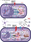
Oxidative damage and mitochondria-associated membranes. (1) Under normal conditions, ER stress activates the UPR which acts to restore homeostasis. Oxidative and hormonal stress stimulate ER-to-Mitochondria Ca2+ transfer (via inositol triphosphate receptors (IP3Rs)). MAM proteins (SIGMAR-1 and DISC1) act to stabilize IP3Rs and limit overall mitochondrial calcium accumulation. Independently, DISC1 acts to counteract corticosterone-induced stress (HPA-axis overactivity) and SIGMAR-1 acts to inhibit pro-inflammatory gene expression (NF-kB) and assist in the formation of mature BDNF. (2) Mitochondrial dysfunction in BD leads to increased oxidative damage, overwhelming the UPR. SIGMAR-1/DISC dysfunction can lead to decreased BDNF expression, loss of feedback on hormonal/oxidative stress signals, and increased IP3R ligand binding (subsequently increasing calcium influx into mitochondria). Risk SNPs associated with the CACNA1C locus can lead to even greater Ca2+ activity, further overwhelming anti-apoptotic signals (bcl-2). (3) Taken together, this dysfunction leads to increased pro-inflammatory signaling, NLRP3 inflammasome assembly at the ER-mitochondrial border, and eventually apoptosis, pyropoptosis, or autophagy. Subsequent release of cellular contents can cause amplification of extracellular inflammatory signaling and neurotoxicity (detailed in Figs. 3 and 4)
Fig. 3
Fig. 3
NL3P3 inflammasome activation. (1) Increased cellular stress causes the release of toxic metabolites (ATP, uric acid, glutamate, DAMPs, PAMPs, and others), increasing influx of extracellular calcium through P2X7, NMDA, and L-type calcium channels (LTCCs). ATP-P2X7 upregulation independently induces neuroinflammation (via NLRP3 activation, NF-kB, NFAT, GSK3β and VEGF signaling—not shown here), increases NMDA-excitotoxicity, and has been associated with rapid cycling in BD. (2) Mitochondrial dysfunction (decreased complex I activity) results in decreased oxidative phosphorylation and increased generation of ROS, Ca2+, as well as oxidized lipids and mtDNA. Intracellular Ca2+ imbalance can overwhelm deficient mitochondrial buffering capacity in BD, leading to fragmentation, morphological abnormalities, and eventually apoptosis. By any means, apoptotic signals (especially release of oxidized mtDNA), which potently upregulate NLRP3 activity [55]. (3) Damage signals activate the NLRP3 inflammasome, which (via caspase-1 cleavage) causes the elaboration of mature IL-1β and IL-18 as well as pore formation in the cell membrane (pyropoptosis), and ultimately the release of these deleterious intracellular molecules into the extracellular space. DAMPs/PAMPs also upregulate proinflammatory gene expression (NF-kB) and subsequent release of IL-1β, IL-6, IL-18, TNF-α, and other cytokines. (4) Once in the extracellular space, these agents act to amplify inflammation, activating surrounding microglia, increasing BBB permeability, recruiting peripheral immune cells, and upregulating the complement cascade (via Hmgb-1 and S100a9—MBL binding), causing sustained, sterile inflammation in the brain. Sublytic membrane attack complex (MAC) stimulation can also lead to increased mitochondrial calcium influx and loss of mitochondrial membrane potential, resulting in NLRP3 activation (not shown here) [54]. (5) In healthy individuals, these signals serve as repair mechanisms, and are deactivated by anti-inflammatory feedback (mainly from microglia and T regulatory cells), restoring homeostasis. In BD, chronic mitochondrial dysfunction and lack of proper feedback signaling results in amplification of inflammation and chronic neurodegeneration
Fig. 4
Fig. 4
The Kynurenic pathway, glutamate, and neuroplasticity. (1) 95% of free, extracellular tryptophan is converted into KYN via IDO and TDO. BD, stress, and metabolic disease augment this conversion, decreasing free serotonin/melatonin with resultant increases in oxidative/inflammatory signaling. (2) Inside astrocytes, KYN is converted into KynA, which exerts a net neuroprotective effect by several mechanisms. (3) Through extra-synaptic NMDA-inhibition, ketamine shunts glutamate towards AMPA receptors and blocks QA influx, ultimately upregulating BDNF-mediated synaptic plasticity, in concert with an increase in KynA/QA ratio (NMDA receptor-kynurenic competition). (4) Inside microglia, KYN is converted to 3-HK and QA which exert various neurotoxic effects. (5) Proinflammatory cytokines increase QA production by increasing IDO activity and shunting tryptophan into microglia. This in turn amplifies the inflammatory signal, causing influx of peripheral immune cells, which exponentiate QA production and subsequent toxicity. (6) Under chronic stress (HPA overactivity), cortisol loses its feedback inhibition on pro-inflammatory signals, and continues to upregulate QA production though TDO/IDO induction and IFN-γ modulation, exacerbating the neurotoxic/inflammatory environment (discussed in Sect. 7). (7) Astrocytic processes form a “cradle” (left side) around synaptic connections and serve as the major site for glutamate reuptake, limiting excess glutamate spillover into extrasynaptic NMDA receptors. Astrocytes also exert negative feedback (not shown) to suppress microglial overactivation. Chronic inflammation, exacerbated by acute mood episodes (right side) disrupts the astrocytic cradle and impairs feedback mechanisms, resulting in extracellular glutamate accumulation and microglial overactivation. Activated microglia do engage in modest glutamate reuptake, but it is suppressed by QA and outpaced by their immune-stimulated release of various toxic metabolites (QA, ROS, Glutamate, cytokines, damaged organelles, etc.). Extrasynaptic NMDA activation suppresses BDNF expression, impairing synaptic homeostasis. Ultimately these signals result in a chronic reciprocal exacerbation of glutamate excitotoxicity, inflammation, and overall neurotoxicity (discussed in Sect. 8)

References

    1. Andrejew R, Oliveira-Giacomelli Á, Ribeiro DE, et al. The P2X7 receptor: central hub of brain diseases. Front Mol Neurosci. 2020;13:124. doi: 10.3389/fnmol.2020.00124. - DOI - PMC - PubMed
    1. SayuriYamagata A, Brietzke E, Rosenblat JD, Kakar R, McIntyre RS. Medical comorbidity in bipolar disorder: the link with metabolic-inflammatory systems. J Affect Disord. 2017;211:99–106. doi: 10.1016/j.jad.2016.12.059. - DOI - PubMed
    1. Perugi G, Quaranta G, Belletti S, et al. General medical conditions in 347 bipolar disorder patients: clinical correlates of metabolic and autoimmune-allergic diseases. J Affect Disord. 2015;170:95–103. doi: 10.1016/j.jad.2014.08.052. - DOI - PubMed
    1. Panaccione I, Spalletta G, Sani G. Neuroinflammation and excitatory symptoms in bipolar disorder. Neuroimmunol Neuroinflam. 2015;2(4):215–227. doi: 10.4103/2347-8659.167304. - DOI
    1. Khairova RA, MacHado-Vieira R, Du J, Manji HK. A potential role for pro-inflammatory cytokines in regulating synaptic plasticity in major depressive disorder. Int J Neuropsychopharmacol. 2009;12(4):561–578. doi: 10.1017/S1461145709009924. - DOI - PMC - PubMed