. 2021 Jun 10;23(1):77.
doi: 10.1186/s12968-021-00752-1.

Demographic, multi-morbidity and genetic impact on myocardial involvement and its recovery from COVID-19: protocol design of COVID-HEART-a UK, multicentre, observational study

Collaborators, Affiliations

Demographic, multi-morbidity and genetic impact on myocardial involvement and its recovery from COVID-19: protocol design of COVID-HEART-a UK, multicentre, observational study

Miroslawa Gorecka et al. J Cardiovasc Magn Reson. .

Abstract

Background: Although coronavirus disease 2019 (COVID-19) is primarily a respiratory illness, myocardial injury is increasingly reported and associated with adverse outcomes. However, the pathophysiology, extent of myocardial injury and clinical significance remains unclear.

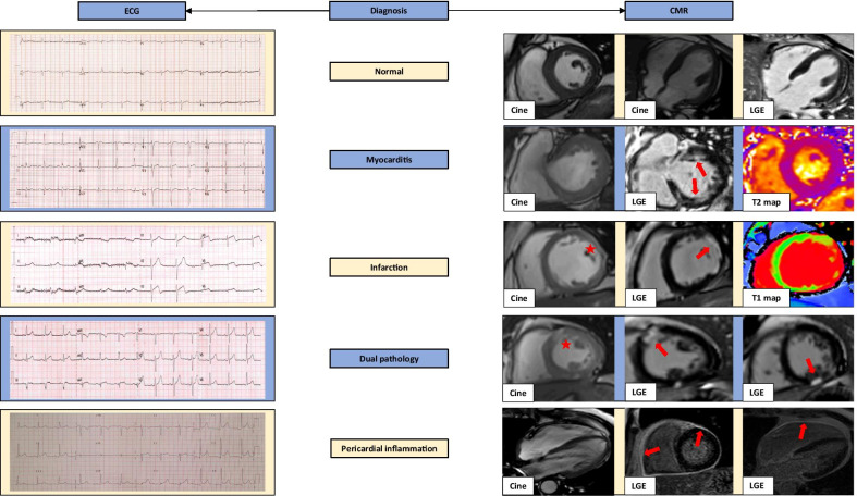
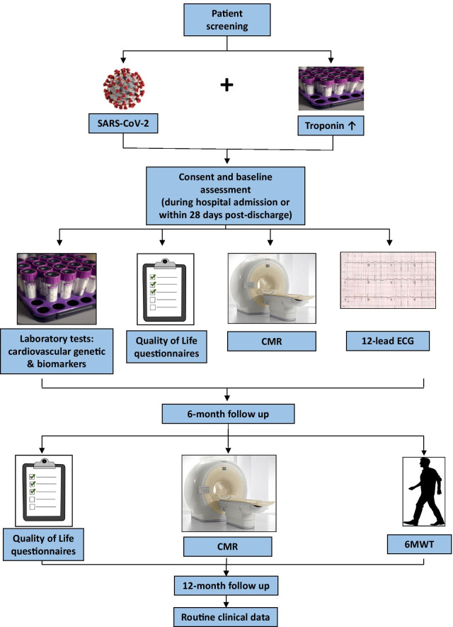
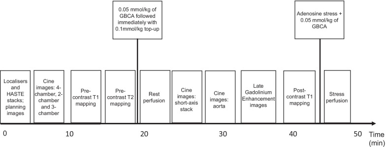
Methods: COVID-HEART is a UK, multicentre, prospective, observational, longitudinal cohort study of patients with confirmed COVID-19 and elevated troponin (sex-specific > 99th centile). Baseline assessment will be whilst recovering in-hospital or recently discharged, and include cardiovascular magnetic resonance (CMR) imaging, quality of life (QoL) assessments, electrocardiogram (ECG), serum biomarkers and genetics. Assessment at 6-months includes repeat CMR, QoL assessments and 6-min walk test (6MWT). The CMR protocol includes cine imaging, T1/T2 mapping, aortic distensibility, late gadolinium enhancement (LGE), and adenosine stress myocardial perfusion imaging in selected patients. The main objectives of the study are to: (1) characterise the extent and nature of myocardial involvement in COVID-19 patients with an elevated troponin, (2) assess how cardiac involvement and clinical outcome associate with recognised risk factors for mortality (age, sex, ethnicity and comorbidities) and genetic factors, (3) evaluate if differences in myocardial recovery at 6 months are dependent on demographics, genetics and comorbidities, (4) understand the impact of recovery status at 6 months on patient-reported QoL and functional capacity.

Discussion: COVID-HEART will provide detailed characterisation of cardiac involvement, and its repair and recovery in relation to comorbidity, genetics, patient-reported QoL measures and functional capacity.

Clinical trial registration: ISRCTN 58667920. Registered 04 August 2020.

Keywords: COVID-19; Cardiovascular disease; Cardiovascular magnetic resonance; Coronavirus; Myocardial infarction; Myocardial inflammation; Myocardial repair; Myocarditis; Myopericarditis; myocardial injury.


Conflict of interest statement

CB is employed by the University of Glasgow which holds research and/or consultancy agreements with AstraZeneca, Abbott Vascular, Boehringer Ingelheim, GSK, HeartFlow, Opsens, and Novartis. CBD is the chief executive officer (part-time) of the Society for Cardiovascular Magnetic Resonance (SCMR). All other authors have no conflicts to declare.

Figures

Fig. 1
Fig. 1
Electrocardiogram (ECG) and cardiovascular magnetic resonance (CMR) examples in troponin positive COVID-19. Potential diagnoses include: no abnormalities detected, myocarditis, infarction, dual pathology and pericardial inflammation. First example shows a normal 12-lead electrocardiogram (ECG) and a normal CMR study. Second example shows myocarditis, with anterolateral ST segment flattening. CMR shows normal function but patchy mid-wall enhancement in the anterior and inferior wall (red arrows) with T2 map showing co-localised oedema. The third example shows myocardial infarction with ECG anterolateral ST changes and thinning of left ventricular (LV) wall predominantly in the anterolateral segment on the short-axis image (red asterisk); corresponding LGE image shows an ischaemic pattern transmural scar (red arrow); T1 maps show elevated T1 values in the lateral wall. The fourth example shows a case of dual pathology due to myocarditis and infarction. The ECG shows hyperacute T-waves in the anterolateral leads and the CMR study shows a short-axis cine with increased signal intensity in the anterior/anteroseptal segment (red asterisk). The two LGE images reveal an ischaemic scar in this area extending from subendocardium as well as an area of sub-epicardial enhancement in the inferior wall consistent with myocarditis (red arrows). The last case presents pericardial inflammation. The ECG shows widespread concave ST-segment elevation. The 4-chamber cine is unremarkable, whereas the corresponding LGE images show pericardial enhancement (red arrows)
Fig. 2
Fig. 2
Study flow diagram. COVID-19 patients, who have an elevated troponin, will be invited to participate in the study. Patients who are recruited undergo multimodality assessment at index visit: either during their hospital admission or within 28 days of discharge. Assessment includes a 12-lead ECG, CMR study, genetic and immunological blood tests, and Quality of Life questionnaires. Patients are subsequently invited for a second visit at 6 months. At this point they undergo a repeat CMR scan, quality of life assessments and 6-min walk test. Further follow up at 12 months will be conducted via examination of routine clinical data, available through general practitioners, electronic hospital records, NICOR and NHS Digital, and in collaboration with other nationally recognised COVID-19 studies
Fig. 3
Fig. 3
COVID-HEART study CMR protocol. The full protocol takes approximately an hour, and comprises (in order of acquisition): localisers and axial HASTE stack, cine images of the left ventricle in long-axis views, pre-contrast T1 mapping and T2 mapping, rest myocardial perfusion after injection of 0.05 mmol/kg of gadolinium based contrast agent (GBCA) followed immediately with 0.1 mmol/kg top-up (giving a total dose of 0.15 mmol/kg). Then a ventricular short-axis stack and aortic cine images (aortic distensibility, followed by late gadolinium enhancement (LGE) imaging and post-contrast T1 mapping. The last component (dependent on patient suitability) is stress perfusion imaging, performed after administration of adenosine stress at dose of 140-210mcg/kg/min, utilising a further 0.05 mmol/kg of GBCA

References

    1. Bandyopadhyay D, Akhtar T, Hajra A, Gupta M, Das A, Chakraborty S, et al. COVID-19 pandemic: cardiovascular complications and future implications. Am J Cardiovasc Drugs. 2020;20(4):311–324. doi: 10.1007/s40256-020-00420-2. - DOI - PMC - PubMed
    1. Gavriatopoulou M, Korompoki E, Fotiou D, Ntanasis-Stathopoulos I, Psaltopoulou T, Kastritis E, et al. Organ-specific manifestations of COVID-19 infection. Clin Exp Med. 2020 doi: 10.1007/s10238-020-00648-x. - DOI - PMC - PubMed
    1. Raman B, Cassar MP, Tunnicliffe EM, Filippini N, Griffanti L, Alfaro-Almagro F, et al. Medium-term effects of SARS-CoV-2 infection on multiple vital organs, exercise capacity, cognition, quality of life and mental health, post-hospital discharge. EClinicalMedicine. 2020;31:100683. doi: 10.1016/j.eclinm.2020.100683. - DOI - PMC - PubMed
    1. Bansal M. Cardiovascular disease and COVID-19. Diabetes Metab Syndr. 2020;14(3):247–250. doi: 10.1016/j.dsx.2020.03.013. - DOI - PMC - PubMed
    1. Aghagoli G, Gallo Marin B, Soliman LB, Sellke FW. Cardiac involvement in COVID-19 patients: risk factors, predictors, and complications: a review. J Cardiac Surg. 2020 doi: 10.1111/jocs.14538. - DOI - PMC - PubMed

Publication types

MeSH terms

Associated data