LncRNA CTD-2528L19.6 prevents the progression of IPF by alleviating fibroblast activation
- PMID: 34112765
- PMCID: PMC8192779
- DOI: 10.1038/s41419-021-03884-5
LncRNA CTD-2528L19.6 prevents the progression of IPF by alleviating fibroblast activation
Abstract
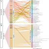
Long non-coding RNAs (lncRNAs) have emerged as critical factors for regulating multiple biological processes during organ fibrosis. However, the mechanism of lncRNAs in idiopathic pulmonary fibrosis (IPF) remains incompletely understood. In the present study, two sets of lncRNAs were defined: IPF pathogenic lncRNAs and IPF progression lncRNAs. IPF pathogenic and progression lncRNAs-mRNAs co-expression networks were constructed to identify essential lncRNAs. Network analysis revealed a key lncRNA CTD-2528L19.6, which was up-regulated in early-stage IPF compared to normal lung tissue, and subsequently down-regulated during advanced-stage IPF. CTD-2528L19.6 was indicated to regulate fibroblast activation in IPF progression by mediating the expression of fibrosis related genes LRRC8C, DDIT4, THBS1, S100A8 and TLR7 et al. Further studies showed that silencing of CTD-2528L19.6 increases the expression of Fn1 and Collagen I both at mRNA and protein levels, promoted the transition of fibroblasts into myofibroblasts and accelerated the migration and proliferation of MRC-5 cells. In contrast, CTD-2528L19.6 overexpression alleviated fibroblast activation in MRC-5 cells induced by TGF-β1. LncRNA CTD-2528L19.6 inhibited fibroblast activation through regulating the expression of LRRC8C in vitro assays. Our results suggest that CTD-2528L19.6 may prevent the progression of IPF from early-stage and alleviate fibroblast activation during the advanced-stage of IPF. Thus, exploring the regulatory effect of lncRNA CTD-2528L19.6 may provide new sights for the prevention and treatment of IPF.
Conflict of interest statement
The authors declare no competing interests.
Figures


Similar articles
-
Interaction of long noncoding RNAs and microRNAs in the pathogenesis of idiopathic pulmonary fibrosis.Physiol Genomics. 2015 Oct;47(10):463-9. doi: 10.1152/physiolgenomics.00064.2015. Epub 2015 Aug 12. Physiol Genomics. 2015. PMID: 26269497 Free PMC article.
-
LncRNA PFAR contributes to fibrogenesis in lung fibroblasts through competitively binding to miR-15a.Biosci Rep. 2019 Jul 18;39(7):BSR20190280. doi: 10.1042/BSR20190280. Print 2019 Jul 31. Biosci Rep. 2019. PMID: 31273058 Free PMC article.
-
Systematic analyses identify the anti-fibrotic role of lncRNA TP53TG1 in IPF.Cell Death Dis. 2022 Jun 4;13(6):525. doi: 10.1038/s41419-022-04975-7. Cell Death Dis. 2022. PMID: 35661695 Free PMC article.
-
Decrypting the crosstalk of noncoding RNAs in the progression of IPF.Mol Biol Rep. 2020 Apr;47(4):3169-3179. doi: 10.1007/s11033-020-05368-9. Epub 2020 Mar 16. Mol Biol Rep. 2020. PMID: 32180083 Review.
-
Wilms Tumor 1-Driven Fibroblast Activation and Subpleural Thickening in Idiopathic Pulmonary Fibrosis.Int J Mol Sci. 2023 Feb 2;24(3):2850. doi: 10.3390/ijms24032850. Int J Mol Sci. 2023. PMID: 36769178 Free PMC article. Review.
Cited by
-
Effect of Hypoxia in the Transcriptomic Profile of Lung Fibroblasts from Idiopathic Pulmonary Fibrosis.Cells. 2022 Sep 27;11(19):3014. doi: 10.3390/cells11193014. Cells. 2022. PMID: 36230977 Free PMC article.
-
Noncoding RNAs: Master Regulator of Fibroblast to Myofibroblast Transition in Fibrosis.Int J Mol Sci. 2023 Jan 16;24(2):1801. doi: 10.3390/ijms24021801. Int J Mol Sci. 2023. PMID: 36675315 Free PMC article. Review.
-
Platyconic acid A‑induced PPM1A upregulation inhibits the proliferation, inflammation and extracellular matrix deposition of TGF‑β1‑induced lung fibroblasts.Mol Med Rep. 2022 Nov;26(5):329. doi: 10.3892/mmr.2022.12845. Epub 2022 Sep 7. Mol Med Rep. 2022. PMID: 36069235 Free PMC article.
-
Baicalein attenuates bleomycin-induced lung fibroblast senescence and lung fibrosis through restoration of Sirt3 expression.Pharm Biol. 2023 Dec;61(1):288-297. doi: 10.1080/13880209.2022.2160767. Pharm Biol. 2023. PMID: 36815239 Free PMC article.
-
Potential function exploration of lncRNAs in idiopathic pulmonary fibrosis: insights from whole transcriptome sequencing data analysis.Clinics (Sao Paulo). 2025 Aug 8;80:100732. doi: 10.1016/j.clinsp.2025.100732. Online ahead of print. Clinics (Sao Paulo). 2025. PMID: 40782499 Free PMC article.
References
Publication types
MeSH terms
Substances
Grants and funding
LinkOut - more resources
Full Text Sources
Other Literature Sources
Miscellaneous

