. 2021 Jun 10;12(1):3514.
doi: 10.1038/s41467-021-23120-0.

Enhanced mechanosensing of cells in synthetic 3D matrix with controlled biophysical dynamics

Affiliations

Enhanced mechanosensing of cells in synthetic 3D matrix with controlled biophysical dynamics

Boguang Yang et al. Nat Commun. .

Abstract

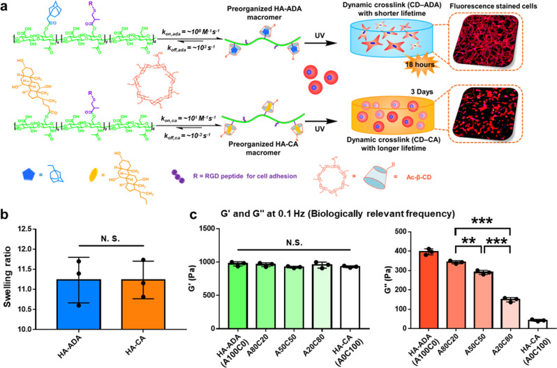
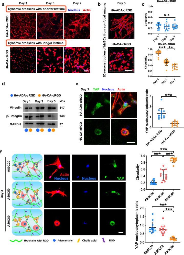
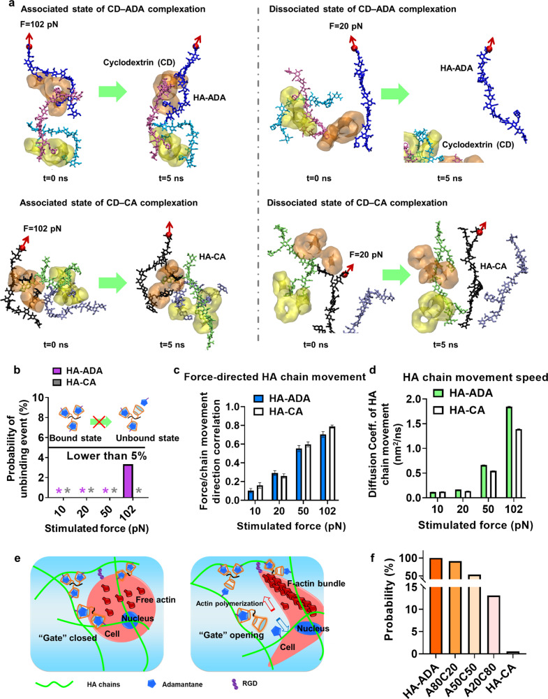
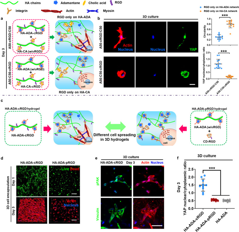
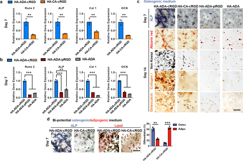
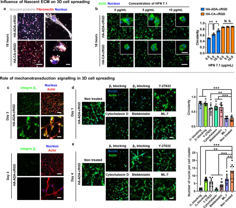
3D culture of cells in designer biomaterial matrices provides a biomimetic cellular microenvironment and can yield critical insights into cellular behaviours not available from conventional 2D cultures. Hydrogels with dynamic properties, achieved by incorporating either degradable structural components or reversible dynamic crosslinks, enable efficient cell adaptation of the matrix and support associated cellular functions. Herein we demonstrate that given similar equilibrium binding constants, hydrogels containing dynamic crosslinks with a large dissociation rate constant enable cell force-induced network reorganization, which results in rapid stellate spreading, assembly, mechanosensing, and differentiation of encapsulated stem cells when compared to similar hydrogels containing dynamic crosslinks with a low dissociation rate constant. Furthermore, the static and precise conjugation of cell adhesive ligands to the hydrogel subnetwork connected by such fast-dissociating crosslinks is also required for ultra-rapid stellate spreading (within 18 h post-encapsulation) and enhanced mechanosensing of stem cells in 3D. This work reveals the correlation between microscopic cell behaviours and the molecular level binding kinetics in hydrogel networks. Our findings provide valuable guidance to the design and evaluation of supramolecular biomaterials with cell-adaptable properties for studying cells in 3D cultures.


Conflict of interest statement

The authors declare no competing interests.

Figures

Fig. 1
Fig. 1. The supramolecular hydrogels stabilized by reversible host–guest crosslinks with different binding kinetics possess differential dynamic properties.
a Schematic illustration of the preparation of supramolecular hyaluronic acid hydrogels stabilized by different pairs of host–guest complexation (cyclodextrin–adamantane/cyclodextrin–cholic acid or CD–ADA/CD–CA) and the monitoring of 3D spreading of encapsulated hMSCs. b The swelling ratio of hydrogels measured after incubating in culture medium for 3 days. Data are presented as mean values ± SD (standard deviation), n = 3 independent hydrogels per group, N.S. indicates no statistical difference. (two-tailed Student’s t-test). c Average value of G′ and G″ from rheological analysis at the frequency of 0.1 Hz and 1% strain. Data are presented as mean values ± SD, n = 3 independent hydrogels per group, N.S. indicates no statistical difference (ANOVA), **p < 0.01, ***p < 0.001 (two-tailed Student’s t-test) (A80C20, A50C50, A20C80 were prepared by mixing HA–ADA and HA–CA at the weight ratio of 80%:20%, 50%:50%, or 20%:80%, respectively).
Fig. 2
Fig. 2. hMSCs exhibit differential spreading within 3D hydrogels stabilized by dynamic crosslinks with varying lifetimes.
a Representative image of hMSCs encapsulated in hydrogels from different groups (HA–ADA–cRGD and HA–CA–cRGD) on culture days 1, 3, and 7 stained for actin (red) and nuclei (blue). Scale bar =100 µm. b 3D reconstruction of hMSCs from confocal images. Scale bar =100 µm. c The circularity of the hMSCs encapsulated within the hydrogels from the different groups (HA–ADA–cRGD and HA–CA–cRGD) (The average cell circularity index was calculated according to C = 4πA/P2, where A is the cell area and P is the cell perimeter). Data are presented as mean values ± SD, n = 12 cells per group from 2 independent hydrogels; N.S. indicates no statistical difference, **p < 0.01, ***p < 0.001 (ANOVA or two-tailed Student’s t-test). d Western blot analysis of vinculin and β1 integrin protein expression in hMSCs after 3 days of osteogenic culture in hydrogels prepared with different lifetime crosslinks. The samples are derived from the same experiment, and gels/blots are processed in parallel. (Data of Relative intensity details are in the Supplementary Table 2) e Representative immunofluorescence staining against F-actin (red), nuclei (blue), and YAP (green) in hMSCs encapsulated in HA–ADA–cRGD and HA–CA–cRGD hydrogels after 3 days of culture (scale bar = 50 μm) and quantification of the nuclear YAP fluorescence intensity (intensity ratio between the nucleus and cytoplasm) Data are presented as mean values ± SD, n = 12 cells per group from two independent hydrogels; ***p < 0.001 (two-tailed Student’s t-test). f Representative immunofluorescence staining against F-actin (red), nuclei (blue), and YAP (green) in hMSCs encapsulated in hydrogels prepared with different weight ratios of HA–ADA and HA–CA guest polymer after 3 days of culture (A80C20, A50C50, A20C80 were prepared by mixing HA–ADA–cRGD and HA–CA–cRGD at the weight ratio of 80%:20%, 50%:50%, or 20%:80%, respectively) and quantification of the cell circularity and nuclear YAP fluorescence intensity (intensity ratio between the nucleus and cytoplasm). Data are presented as mean values ± SD, n = 12 cells per group from two independent hydrogels, ***p < 0.001 (two-tailed Student’s t-test), Scale bar = 25 μm.
Fig. 3
Fig. 3. MD and KMC simulation results verify the key role of binding kinetics of host–guest crosslinks in enabling cell-mediated hydrogel network reorganization.
a MD simulation snapshots showing an application of a force on one end (red dot) of the HA segment when the host–guest crosslink in HA–ADA or HA–CA hydrogels is in the bound and the unbound state, respectively. Two oligomerized acryloyl β-cyclodextrin crosslinkers (each contains three CDs) are shown in orange and yellow, respectively. b For a given magnitude of applied force, sixty 5-ns simulations were performed with CDADA pairs in the bound state. Among the total 240 simulations, unbinding events were only observed in two simulations with F = 102 pN. * means that the value is 0. For the CDCA pairs in the bound state, 60 simulations performed at F = 102 pN revealed no unbinding event. c In a separate set of 480 5-ns simulations with a given CDADA or CDCA pair initially in the unbound state, we measured the correlation coefficient (value ranges from −1 to 1) between the direction of the applied force and the movement of the corresponding HA chain. Error bars represent the standard error of the mean. n = 60. d In the same set of simulations shown in c, the “speed” of HA chain movement is characterized by the diffusion coefficient of the guest molecule closest to the HA end being pulled. The sixty simulations performed at a given force are combined to estimate the diffusion coefficient from a linear regression model, the error of which is smaller than the line width (Supplementary Table 3). e Schematics of parallel actin bundles in filopodia and the crosslinked HA network they face. f Probability for a gate-opening event to occur within the estimated timescale of actin polymerization (~0.01 s) at F = 10 pN based on sets of 100,000 KMC calculations assuming n = 2 in acrylated host complexes (see Supplementary Information for details).
Fig. 4
Fig. 4. The precise and covalent conjugation of cell-adhesive ligands to the hydrogel subnetwork connected by crosslinks with fast binding kinetics is required for efficient 3D spreading and mechanosensing of hMSCs.
a Schematic illustration of the preparation of A50C50 hydrogels with selective RGD conjugations and 3D cell culture in the hydrogels. RGD peptide was only conjugated either to the HA–ADA subnetwork (A50-cRGD:C50) or to HA–CA subnetwork (A50:C50-cRGD) in the A50C50 hydrogels. b Representative immunofluorescence staining against F-actin (red), nuclei (blue), and YAP (green) in hMSCs encapsulated in A50C50 hydrogels with selective RGD conjugations after 3 days of culture (scale bar = 25 μm) and quantification of the circularity and nuclear YAP fluorescence intensity (intensity ratio between the nucleus and cytoplasm). Data are presented as mean values ± SD, n = 12 cells per group from two independent hydrogels; ***p < 0.001 (two-tailed Student’s t-test). c Schematic illustration of the preparation of hydrogels with different RGD conjugation methods (HA–ADA–cRGD and HA–ADA–pRGD) and 3D cell culture with different RGD conjugation methods. d Representative images of hMSCs encapsulated within hydrogels (3D cell encapsulation) with different RGD conjugation methods (HA–ADA–cRGD and HA–ADA–pRGD) after 1 and 3 days of culture. Scale bar = 200 µm. e Representative immunofluorescence staining against F-actin (red), nuclei (blue) and pFAK or vinculin (green) in hMSCs cultured in highly dynamic HA–ADA–cRGD hydrogels for 3 days. Scale bar = 50 μm. f Quantification of the nuclear YAP fluorescence intensity (intensity ratio between the nucleus and cytoplasm) in hMSCs encapsulated in hydrogels with different RGD conjugation methods (HA–ADA–cRGD, HA–ADA–pRGD, and HA–ADA) on after 3 days of culture. Data are presented as mean values ± SD, n = 10 cells per group from two independent hydrogels; ***p < 0.001 (two-tailed Student’s t-test).
Fig. 5
Fig. 5. Supramolecular hydrogels with short crosslink lifetime promote osteogenic differentiation of encapsulated hMSCs.
a Quantification of Runx 2, ALP, type I collagen, and OCN gene expression of hMSCs encapsulated in the hydrogels (HA–ADA–cRGD and HA–CA–cRGD) by RT-PCR after 7 days of osteogenic culture. Values are normalized to expression levels within HA–ADA–cRGD. Data are presented as mean values ± SD, n = 3 independent hydrogels; **p < 0.01 (two-tailed Student’s t-test). b Quantification of Runx 2, ALP, type I collagen, and OCN gene expression of hMSCs encapsulated in the hydrogels (HA–ADA–cRGD, HA–ADA–pRGD, and HA–ADA) by RT-PCR after 7 days of osteogenic culture. Values are normalized to expression levels within HA–ADA–cRGD; Data are presented as mean values ± SD, n = 3 independent hydrogels; **p < 0.01, ***p < 0.001 (two-tailed Student’s t-test). c ALP staining (Fast Blue; osteogenic biomarker, blue) of hMSC-laden hydrogels of different groups after 7 days of osteogenic culture, and Alizarin red staining, Von Kossa staining, and type I collagen and OCN immunohistochemical staining of hMSC-laden hydrogels of different groups after 14 days of osteogenic culture (scale bar = 100 μm). d Representative ALP and lipid staining and percentage differentiation of hMSCs within HA–ADA–cRGD and HA–CA–cRGD hydrogels following 7 days mixed osteogenic/adipogenic-media incubation (Black arrow pointing to ALP (osteogenesis) or lipid-containing cells) (Scale bar = 100 μm). Data are presented as mean values ± SD, n = 3 independent hydrogels; **p < 0.01, ***p < 0.001 (two-tailed Student’s t-test).
Fig. 6
Fig. 6. The ultra-rapid cell spreading and aggregation in HA–ADA–cRGD hydrogels is regulated by cell–nascent ECM interaction, cell adhesion structures containing β1 class integrins, and actomyosin-based contractility.
a Representative images (magnifications on the right) of nascent proteins (white) and fibronectin (red) secreted by hMSCs encapsulated in HA–ADA–cRGD and HA–CA–cRGD gels within 18 h (scale bars, 200 µm (pictures on the left) and 20 μm (pictures on the right)). b Representative images (scale bars, 200 µm (main image) and 10 µm (inset)) of F-actin (green) expressed by the cells encapsulated in HA–ADA–cRGD and HA–CA–cRGD hydrogels after 18 h of treatment with different concentrations of a monoclonal antibody against the cell-adhesive domain of human fibronectin (HFN 7.1, 0, 5, 10 µg/mL, see Supplementary Fig. 19 for cell viability). Circularity of hMSCs encapsulated in HA–ADA–cRGD and HA–CA–cRGD hydrogels after 18 h of treatment with different concentrations of HFN 7.1 (0, 5, 10 µg/mL). Data are presented as mean values ± SD, n = 3 independent hydrogels, *p < 0.05, **p < 0.01 (two-tailed Student’s t-test). c Representative immunofluorescence staining against F-actin (red), nuclei (blue), and β1 class integrins or β3 class integrins (green) in hMSCs cultured in highly dynamic HA–ADA–cRGD hydrogels for 3 days (images on the top: scale bar = 200 μm. Images on the bottom: scale bar = 50 μm). d Cell spreading in the highly dynamic HA–ADA–cRGD hydrogels after 1 day of culture with or without treatment with integrin-blocking antibodies, a myosin inhibitor (blebbistatin), a myosin light chain kinase inhibitor (ML-7), a ROCK inhibitor (Y-27632), or an inhibitor of actin polymerization (Cytochalasin D). Scale bar = 100 μm. The circularity of the hMSCs encapsulated within the hydrogels treated with different inhibitors. (The average circularity value is calculated according to C = 4πA/P2, where A is the area occupied by the cell and P is the perimeter of the cell). Data are presented as mean values ± SD, n = 10 cells per group from two independent hydrogels; **p < 0.01, ***p < 0.001 (two-tailed Student’s t-test). e Multi-cell assembly in the highly dynamic HA–ADA–cRGD hydrogels after 4 days of culture with or without treatment with blocking antibodies and inhibitors. Scale bar = 100 μm. The quantification of the multicellularity of the cell clusters within the hydrogels treated with different inhibitors. Data are presented as mean values ± SD, n = 10 cell clusters per group from two independent hydrogels; **p < 0.01, ***p < 0.001 (two-tailed Student’s t-test).

References

    1. Briquez, P. S., Clegg, L. E., Martino, M. M., Mac Gabhann, F. & Hubbell, J. A. Design principles for therapeutic angiogenic materials. Nat. Rev. Mater.1, 15006 (2016).
    1. Even-Ram S, Artym V, Yamada KM. Matrix control of stem cell fate. Cell. 2006;126:645–647. doi: 10.1016/j.cell.2006.08.008. - DOI - PubMed
    1. Engler AJ, Sen S, Sweeney HL, Discher DE. Matrix elasticity directs stem cell lineage specification. Cell. 2006;126:677–689. doi: 10.1016/j.cell.2006.06.044. - DOI - PubMed
    1. Hoffman BD, Grashoff C, Schwartz MA. Dynamic molecular processes mediate cellular mechanotransduction. Nature. 2011;475:316–323. doi: 10.1038/nature10316. - DOI - PMC - PubMed
    1. Rosales, A. M. & Anseth, K. S. The design of reversible hydrogels to capture extracellular matrix dynamics. Nat. Rev. Mater.1, 15012 (2016). - PMC - PubMed

Publication types

MeSH terms