. 2021 Jun 10;12(1):3534.
doi: 10.1038/s41467-021-23886-3.

Viral infiltration of pancreatic islets in patients with COVID-19

Affiliations

Viral infiltration of pancreatic islets in patients with COVID-19

Charlotte Steenblock et al. Nat Commun. .

Abstract

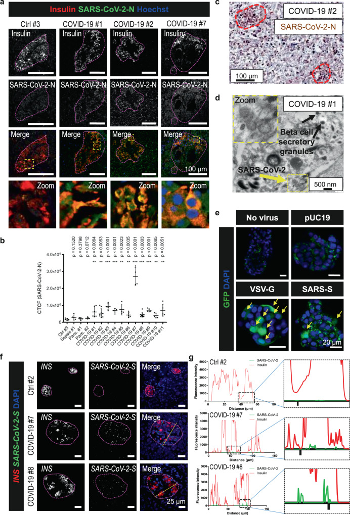
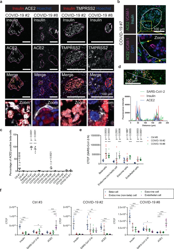
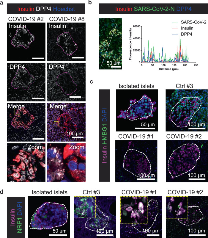
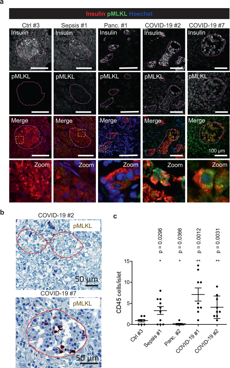
Metabolic diseases are associated with an increased risk of severe COVID-19 and conversely, new-onset hyperglycemia and complications of preexisting diabetes have been observed in COVID-19 patients. Here, we performed a comprehensive analysis of pancreatic autopsy tissue from COVID-19 patients using immunofluorescence, immunohistochemistry, RNA scope and electron microscopy and detected SARS-CoV-2 viral infiltration of beta-cells in all patients. Using SARS-CoV-2 pseudoviruses, we confirmed that isolated human islet cells are permissive to infection. In eleven COVID-19 patients, we examined the expression of ACE2, TMPRSS and other receptors and factors, such as DPP4, HMBG1 and NRP1, that might facilitate virus entry. Whereas 70% of the COVID-19 patients expressed ACE2 in the vasculature, only 30% displayed ACE2-expression in beta-cells. Even in the absence of manifest new-onset diabetes, necroptotic cell death, immune cell infiltration and SARS-CoV-2 viral infection of pancreatic beta-cells may contribute to varying degrees of metabolic dysregulation in patients with COVID-19.


Conflict of interest statement

The authors declare no competing interests.

Figures

Fig. 1
Fig. 1. SARS-CoV-2 infection leads to viral infiltration in the pancreas.
a Immunostainings of pancreas sections from control and COVID-19 patients using antibodies against insulin and SARS-CoV-2-N (sc65653). Representative images from three independent experiments are shown (n = 3). Islets are indicated. Scale bars, 100 µm. b Dot blots of SARS-CoV-2 cellular fluorescence in beta-cells measured on one (n = 1) of three independent stainings in a. Quantification of calculated total cellular fluorescence (CTCF) was performed on n ≥ 5 cells from n ≥ 2 islets per patient. Data were analyzed by unpaired two-sided t-test by comparison with Ctrl #3. Data are presented as mean ± SD. p values are indicated. **p < 0.01; ***p < 0.001. c Immunohistochemistry of pancreas tissue from patient #2 using a different SARS-CoV-2-N antibody (GTX135361) (n = 1). Islets are indicated. Scale bar, 100 µm. d Electron microscopy image of pancreas tissue from patient #1 showing virus-like particles. Two independent experiments with similar results were performed. Scale bar, 500 nm. e Representative images of isolated human islets infected with VSV-G or SARS-CoV-2 pseudoviruses expressing GFP. Results shown are from one of two independent biological experiments (n = 2); each with three technical replicates (n = 3). pUC19 empty vector. Scale bars, 20 µm. Infected cells are indicated with yellow arrows. f Representative images of RNA fluorescence in situ hybridization performed using the RNA scope platform with probes targeting INS and SARS-CoV-2-S (n = 2 independent experiments). Probes against the bacterial dapB gene were used as negative control. Islets are indicated. Scale bars, 25 µm. g Morphometric analysis of SARS-CoV-2-S and INS RNA in situ hybridizations. The fluorescence intensities in the different fluorescence channels along the yellow line were measured.
Fig. 2
Fig. 2. SARS-CoV-2 receptors are expressed in the human pancreas.
a Pancreas sections from COVID-19 patients were immunostained for insulin to mark beta-cells. Additionally, double stainings for ACE2 and TMPRSS2 were performed. Representative images from three independent experiments (n = 3) are shown. Islets are indicated. Scale bars, 100 µm. b Triple immunostaining for ACE2, insulin and the endothelial marker VCAM-1. Representative image from two independent experiments (n = 2) are shown. Islets are indicated. Scale bar, 100 µm. c Dot blot showing the percentage of beta-cells positive for ACE2 in control and COVID-19 patients. All insulin-positive cells in n ≥ 3 islets per patient were counted from three independent experiments (n = 3). Data were analyzed by unpaired two-sided t-test by comparison with Ctrl #3. Data are presented as mean ± SD. d Morphometric analysis of an ACE2/Insulin/SARS-CoV-2-N staining of pancreatic tissue from COVID-19 patient #2 (n = 1). The fluorescence intensities in the indicated fluorescence channels along the white line were measured. Scale bar, 100 µm. e Calculated total cellular fluorescence (CTCF) of SARS-CoV-2 in insulin-positive and -negative islet cells, exocrine cells, and endothelial cells. Data were analyzed by unpaired two-sided t-test by comparison with Ctrl #3. Data are presented as mean ± SD. f CTCF of ACE2 and SARS-CoV-2 was calculated in insulin-positive and -negative islet cells, exocrine cells and endothelial cells. Data were analyzed by two-way ANOVA and Bonferroni posttest. Data are presented as mean ± SD. *p < 0.05; **p < 0.01; ***p < 0.001. In e and f, quantification of CTCF was calculated based on n ≥ 5 cells from n ≥ 2 islets per patient from one (n = 1) out of three independent experiments.
Fig. 3
Fig. 3. Alternative receptors and factors potentially facilitating virus entry.
a Pancreas sections from control patients and deceased COVID-19 patients were immunostained for insulin to mark beta-cells. Additionally, double stainings for DPP4 were carried out. Representative images from two independent experiments (n = 2) are shown. Islets are indicated. Scale bars, 100 µm. b Morphometric analysis of a DPP4/Insulin/SARS-CoV-2-N staining of pancreatic tissue from COVID-19 patient #2 (n = 1). The fluorescence intensities in the indicated fluorescence channels along the white line were measured. Scale bar, 50 µm. c Double stainings for insulin and HMBG1. Representative images from two independent experiments (n = 2) are shown. Islets are indicated. Scale bars, 50 µm for isolated islets, otherwise 100 µm. d Double stainings for insulin and NRP1. Representative images from two independent experiments (n = 2) are shown. Islets are indicated. Scale bars, 50 µm for isolated islets, otherwise 100 µm.
Fig. 4
Fig. 4. SARS-CoV-2 infection leads to necroptosis and immune cell infiltration in the pancreas.
a Immunostainings of pancreas sections from non-COVID-19 patients (control, septic, and with pancreatitis) and from COVID-19 patients using antibodies against insulin and pMLKL marking necroptotic cells. Representative images from three independent experiments (n = 3) are shown. Scale bars, 100 µm. b Immunohistochemistry of pancreas tissue from patient #2 and #7 marking pMLKL-positive cells (n = 1). Islets are indicated. Scale bars, 50 µm. c On immunostainings of pancreas sections from non-COVID-19 patients and from COVID-19 patients stained against insulin and CD45 marking hematopoietic cells, the amount of CD45-positive cells per islet were quantified in n ≥ 7 islets per patient from two independent experiments (n = 2). Data were analyzed by unpaired two-sided t-test by comparison with Ctrl #3. Data in dot plot are presented as mean ± SD. p-values are indicated. *p < 0.05; **p < 0.01.

References

    1. Bornstein SR, Dalan R, Hopkins D, Mingrone G, Boehm BO. Endocrine and metabolic link to coronavirus infection. Nat. Rev. Endocrinol. 2020;16:297–298. doi: 10.1038/s41574-020-0353-9. - DOI - PMC - PubMed
    1. Pal R, Banerjee M. COVID-19 and the endocrine system: exploring the unexplored. J. Endocrinol. Invest. 2020;43:1027–1031. doi: 10.1007/s40618-020-01276-8. - DOI - PMC - PubMed
    1. Somasundaram NP, et al. The impact of SARS-Cov-2 virus infection on the endocrine system. J. Endocr. Soc. 2020;4:bvaa082. doi: 10.1210/jendso/bvaa082. - DOI - PMC - PubMed
    1. Steenblock C, et al. Severe acute respiratory syndrome coronavirus 2 (SARS-CoV-2) and the neuroendocrine stress axis. Mol. Psychiatry. 2020;25:1611–1617. doi: 10.1038/s41380-020-0758-9. - DOI - PMC - PubMed
    1. Alsadhan I, et al. Diabetic ketoacidosis precipitated by Coronavirus disease 2019 infection: case series. Curr. Ther. Res. Clin. Exp. 2020;93:100609. doi: 10.1016/j.curtheres.2020.100609. - DOI - PMC - PubMed

Publication types

MeSH terms