. 2021 Jun 10;11(1):12330.
doi: 10.1038/s41598-021-91300-5.

High-throughput quantitation of SARS-CoV-2 antibodies in a single-dilution homogeneous assay

Affiliations

High-throughput quantitation of SARS-CoV-2 antibodies in a single-dilution homogeneous assay

Markus H Kainulainen et al. Sci Rep. .

Abstract

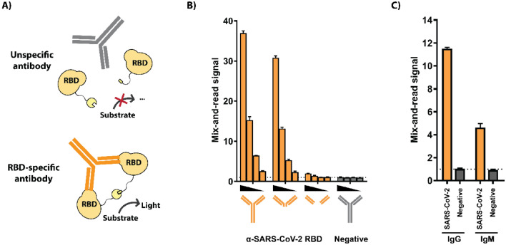
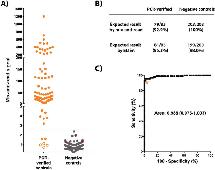
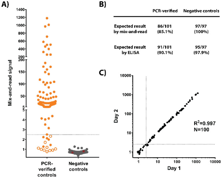
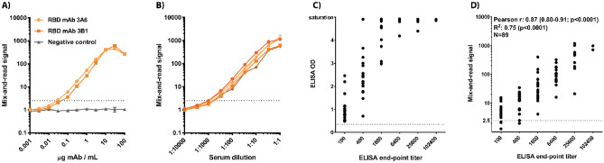
SARS-CoV-2 emerged in late 2019 and has since spread around the world, causing a pandemic of the respiratory disease COVID-19. Detecting antibodies against the virus is an essential tool for tracking infections and developing vaccines. Such tests, primarily utilizing the enzyme-linked immunosorbent assay (ELISA) principle, can be either qualitative (reporting positive/negative results) or quantitative (reporting a value representing the quantity of specific antibodies). Quantitation is vital for determining stability or decline of antibody titers in convalescence, efficacy of different vaccination regimens, and detection of asymptomatic infections. Quantitation typically requires two-step ELISA testing, in which samples are first screened in a qualitative assay and positive samples are subsequently analyzed as a dilution series. To overcome the throughput limitations of this approach, we developed a simpler and faster system that is highly automatable and achieves quantitation in a single-dilution screening format with sensitivity and specificity comparable to those of ELISA.


Conflict of interest statement

The authors declare no competing interests.

Figures

Figure 1
Figure 1
Assay principle. (A) Schematic. Small and large fragments of the NanoBit split luciferase are tethered to SARS-CoV-2 receptor-binding domains (RBD) via peptide linkers. When a specific antibody binds 2 RBD antigens, it forces the luciferase halves into proximity with each other, thereby activating the enzyme. Signal is quantified from a homogeneous assay setup after adding luciferase substrate. (B) Demonstration of assay principle. Dose-responsive assay activation by an RBD-specific monoclonal antibody or corresponding F(ab′)2 or Fab fragments (orange) or control antibody (gray). (C) Activation by specific IgG and IgM antibodies. Human IgG and IgM fractions were purified from a SARS-CoV-2 specific serum pool or a negative control pool and tested for activity in the assay. (B) and (C): technical triplicates with averages and standard deviations presented.
Figure 2
Figure 2
(A) Training set data. Serum samples from cases with earlier nucleic acid confirmed SARS-CoV-2 infection and samples considered true negatives (no COVID-19 diagnosis, or sample collected prior to SARS-CoV-2 emergence) were analyzed by the mix-and-read assay and by RBD ELISA. Out of 85 positive controls, 79 were above proposed cut-offs on both ELISA and mix-and-read assays (orange symbols above signal 2.5), 4 gave no signal distinguishable from negative controls on either assay (open orange symbols), and 2 were positive on ELISA only (orange symbol below 2.5). All negative control signals (N = 203) were below proposed mix-and-read cut-off (gray symbols), but 4 of those samples were positive by ELISA (gray symbols highlighted by red asterisks). (B) Summary of the training set data. (C) Receiver operating characteristics (ROC) analysis of the data and cut-off selection. Cut-off value of 2.5 (as fold over blank samples) was chosen by ROC analysis of the training set data so that 100% specificity could be maintained.
Figure 3
Figure 3
Independent validation set data. (A) Serum samples from confirmed COVID-19 cases and samples considered true negatives were analyzed by RBD ELISA and by the mix-and-read assay using cut-off values determined with the training set. Out of 101 samples with positive SARS-CoV-2 history, 86 were found positive by the mix-and-read assay (orange symbols above signal 2.5), 10 were negative on both assays (open orange symbols), and 5 were positive on ELISA only (orange symbol below 2.5). All negative controls (N = 97, all but one collected before the COVID-19 outbreak) were negative on the mix-and-read assay (gray symbols), while 2 of those samples were positive on ELISA (gray symbols highlighted by red asterisks). (B) Summary of the independent set results. (C) Day-to-day reproducibility of the mix-and-read assay. Samples from the independent validation set were tested on 2 consecutive days and correlation analysis performed on log-transformed values. Samples that gave a discordant qualitative result between the days (near the cut-off, above one day and below the other) were judged negative for purposes of sensitivity and specificity calculations.
Figure 4
Figure 4
Quantitation. (A) Two positive control IgG mAbs and a negative control mAb were titrated to determine the linear range of the mix-and-read signal. (B) Five serum samples that gave a strong signal in initial testing (503–1179 fold over background) were titrated in negative human serum to determine if they reached the saturation level identified by mAbs. (C) Correlation of ELISA end-point titer and ELISA optical density from 1:100 sample dilution. (D) Correlation of ELISA end-point titer and mix-and-read signal. Correlation analysis (Pearson r and 95% confidence interval) and linear regression were performed on log-transformed mix-and-read data and log2-transformed ELISA end-point titers. (A) and (B): technical duplicates with averages and ranges presented.

References

    1. Lerner AM, et al. The COVID-19 serology studies workshop: recommendations and challenges. Immunity. 2020;53:1–5. doi: 10.1016/j.immuni.2020.06.012. - DOI - PMC - PubMed
    1. Wu F, et al. A new coronavirus associated with human respiratory disease in China. Nature. 2020;579:265–269. doi: 10.1038/s41586-020-2008-3. - DOI - PMC - PubMed
    1. WHO. WHO Statement Regarding Cluster of Pneumonia Cases In Wuhan, China. Available at: https://www.who.int/china/news/detail/09-01-2020-who-statement-regarding... (2020).
    1. Yong SEF, et al. Connecting clusters of COVID-19: an epidemiological and serological investigation. Lancet Infect. Dis. 2020;20:809–815. doi: 10.1016/S1473-3099(20)30273-5. - DOI - PMC - PubMed
    1. Long Q-X, et al. Clinical and immunological assessment of asymptomatic SARS-CoV-2 infections. Nat. Med. 2020;26:1200–1204. doi: 10.1038/s41591-020-0965-6. - DOI - PubMed

Publication types