. 2021 Jun 10;11(1):12321.
doi: 10.1038/s41598-021-91830-y.

Facile method for delivering chikungunya viral replicons into mosquitoes and mammalian cells

Affiliations

Facile method for delivering chikungunya viral replicons into mosquitoes and mammalian cells

Hui-Chung Lin et al. Sci Rep. .

Abstract

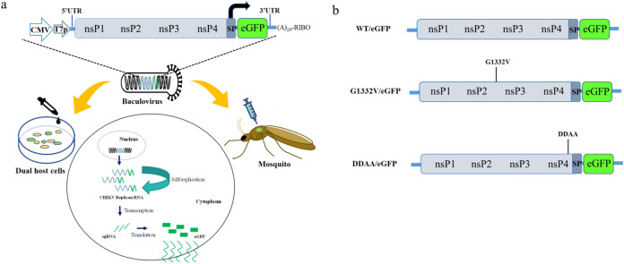
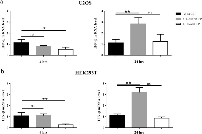
Reverse genetics is an important tool in the elucidation of viral replication and the development of countermeasures; however, these methods are impeded by laborious and inefficient replicon delivery methods. This paper demonstrates the use of a baculovirus to facilitate the efficient delivery of autonomous CHIKV replicons into mosquito and mammalian cells in vitro as well as adult mosquitoes in vivo. The efficacy of this approach was verified via co-localization among an eGFP reporter, nsP1, and dsRNA as well as through the inhibition of an RNA-dependent RNA polymerase (RdRp) null mutation (DDAA) in nsP4, or the treatment of a known antiviral compound (6-azauridine). We also investigated the correlation between CHIKV replicon-launched eGFP expression and the effectiveness of CHIKV replicon variants in inducing IFN-β expression in human cell lines. This delivery method based on a single vector is applicable to mosquito and mammalian cells in seeking to decipher the mechanisms underlying CHIKV replication, elucidate virus-host interactions, and develop antivirals. This study presents an effective alternative to overcome many of the technological issues related to the study and utilization of autonomous arbovirus replicons.


Conflict of interest statement

The authors declare no competing interests.

Figures

Figure 1
Figure 1
Baculovirus vector used to shuttle CHIKV replicons into mosquito and mammal cells: (a) Schematic illustration showing CHIKV replication cycle in mosquito and mammal cells using a single baculovirus. A recombinant baculovirus bearing a DNA cassette of CHIKV replicon-eGFP under the control of a CMV promoter. The baculovirus efficiently delivered CHIKV replicon-eGFP DNA into the nuclei of mosquitoes and mammal cells and adult mosquitoes to induce the expression of eGFP via transduction. T7p, T7 polymerase promoter; 5′ UTR, 5′ untranslated region of CHIKV genome; nsP1–4, non-structural proteins 1–4; SP, subgenomic promoter; 3′ UTR, 3′ untranslated region of CHIKV genome; (A)29, 29 A residues; RIBO, ribozyme site; sgRNA, subgenomic RNA. (b) Schematic diagram showing CHIKV replicon/eGFP constructs. WT/eGFP is a CHIKV replicon derived from LR2006_OPY1strain; G1332V/eGFP, WT/eGFP mutated penultimate glycine residue (P2 residue of 2/3 site) to valine of nsP2; DDAA/eGFP, WT/eGFP mutated the polymerase active site motif Gly-Asp-Asp to Gly-Ala-Ala of nsP4.
Figure 2
Figure 2
Autonomous CHIKV replicon-mediated expression of eGFP in baculovirus transduced into mosquito cell lines, AP-61 (a) or C6/36; (b) cells were transduced using WT/eGFP at indicated MOIs of 0, 1, 5 or 10 in triplicate. At 2 dpt, eGFP-expressing cells were photographed using green (upper panels) or bright field (lower panels) filters. Expression levels of eGFP were quantified (right part). Error bars represent the standard deviation.
Figure 3
Figure 3
Autonomous CHIKV replicon-mediated expression of eGFP via baculovirus transduction in mammalian cell lines, U2OS (a) or HEK293T (b) cells via transduction with WT/eGFP at indicated MOIs of 0, 1, 5 or 10 in triplicate. At 2 dpt, eGFP-expressing cells were photographed using green (upper panels) or bright field (lower panels) filters. Expression levels of eGFP were quantified (right part). Error bars represent the standard deviation.
Figure 4
Figure 4
Characterization of autonomous CHIKV replicon using IFA. AP-61cells were transduced with W/eGFP at MOI of 0.5. At 4 dpt, cells were fixed and co-stained with Mab anti-eGFP antibodies, rabbit anti-CHIKV nsP1 serum, and Hoechst 33342 (upper panels), or co-stained with Mab anti-dsRNA, rabbit anti-CHIKV nsP1 serum, and Hoechst 33342 (lower panels). Merged figures of the three are displayed in the right panels.
Figure 5
Figure 5
Effect of 6-azaudine in inhibiting CHIKV replicon-mediated expression of eGFP in dual host cell lines. U2OS cells (a) were transduced in triplicate using either WT/eGFP or CMV-GFP as a negative control, respectively at an MOI of 5 or 1. AP-61 cells (b) were transduced in triplicate using WT/eGFP at an MOI of 1. Mock or transduced cells growth within a range of 6-AU (0–10 μg/ml) over a period of 24 h. eGFP expression and cell viability were quantified using eGFP or MTT. Error bars indicate standard deviation. Data were combined from three independent experiments.
Figure 6
Figure 6
Autonomous CHIKV replicon-mediated eGFP expression of replicon variants in mosquito cell lines. AP-61 (a) or C6/36 (b) cells were transduced in triplicate using either WT/eGFP, G1332V/eGFP, or DDAA/eGFP at indicated MOIs of 2. At 2 dpt, eGFP-expressing cells were photographed using green (upper panels) or bright field (lower panels) filters. Expression levels of eGFP were quantified (right part). Error bars represent the standard deviation. The statistical significance between WT/eGFP and G1332V/eGFP was analyzed using the Student’s t-test (ns, not significant and ∗∗∗∗p < 0.0001).
Figure 7
Figure 7
Autonomous CHIKV replicon-mediated eGFP expression of replicon variants in mammalian cell lines. U2OS (a) or HEK293T (b) cells were transduced in triplicate using either WT/eGFP, G1332V/eGFP, or DDAA/eGFP at an indicated MOI of 10. At 2 dpt, eGFP-expressing cells were photographed using green (upper panels) or bright field (lower panels) filters. Expression levels of eGFP were quantified (right part). Error bars represent the standard deviation. The statistical significance between WT/eGFP and G1332V/eGFP was analyzed using a Student’s t-test (**p < 0.01 and ∗∗∗∗p < 0.0001).
Figure 8
Figure 8
Time course of CHIKV replicon-mediated eGFP expression of replicon variants in mosquito cell lines (a, AP-61 and C6/36) or mammalian cell lines (b, U2OS and HEK293T) following transduction in triplicate using a recombinant baculovirus at an MOI of 2 or 10, respectively. Expression levels of eGFP were quantified at 1, 2, 3, 5, and 7 dpt. Error bars represent the standard deviation.
Figure 9
Figure 9
Induction of human IFN-β mRNA by CHIKV replicon variants. U2OS (a) or HEK293T (b) cells were transduced in triplicate using either WT/eGFP, G1332V/eGFP, or DDAA/eGFP at an MOI of 10. After incubation for 4 h (left panels) or 24 h (right panels), total RNA was harvested and subjected to qRT-PCR for quantification of IFN-β mRNA expression and normalized to HPRT. Error bars represent the standard deviation. Statistical significance was analyzed using Student’s t-test (ns, not significant; *p < 0.05 and **p < 0.01).
Figure 10
Figure 10
In vivo functional analysis of CHIKV replicon variants in mosquitoes. Adults of A. aegypti (upper panels) and A. albopictus (lower panels) were intrathoracically injected with either WT/eGFP (left panels), G1332V/eGFP (middle panels), or DDAA/eGFP (right panels) at a dose of 5 × 106 pfu/mosquito. The eGFP expression (indicated by white arrows) was photographed (a) and quantified (b). Statistical significance was analyzed using Student’s t-test (*p < 0.05 and **p < 0.01).

Similar articles

Cited by

References

    1. Strauss JH, Strauss EG. The alphaviruses: gene expression, replication, and evolution. Microbiol. Rev. 1994;58:491–562. doi: 10.1128/MR.58.3.491-562.1994. - DOI - PMC - PubMed
    1. Silva LA, Dermody TS. Chikungunya virus: epidemiology, replication, disease mechanisms, and prospective intervention strategies. J. Clin. Investig. 2017;127:737–749. doi: 10.1172/JCI84417. - DOI - PMC - PubMed
    1. Powers AM, Logue CH. Changing patterns of chikungunya virus: re-emergence of a zoonotic arbovirus. J. Gen. Virol. 2007;88:2363–2377. doi: 10.1099/vir.0.82858-0. - DOI - PubMed
    1. Higgs S, Vanlandingham D. Chikungunya virus and its mosquito vectors. Vector Borne Zoonotic Dis. Larchmont NY. 2015;15:231–240. doi: 10.1089/vbz.2014.1745. - DOI - PubMed
    1. Weaver SC, Lecuit M. Chikungunya virus and the global spread of a mosquito-borne disease. N. Engl. J. Med. 2015;372:1231–1239. doi: 10.1056/NEJMra1406035. - DOI - PubMed

Publication types