. 2021 Oct;42(10):1690-1702.
doi: 10.1038/s41401-021-00700-w. Epub 2021 Jun 10.

ATF3 contributes to brucine-triggered glioma cell ferroptosis via promotion of hydrogen peroxide and iron

Affiliations

ATF3 contributes to brucine-triggered glioma cell ferroptosis via promotion of hydrogen peroxide and iron

Shan Lu et al. Acta Pharmacol Sin. 2021 Oct.

Abstract

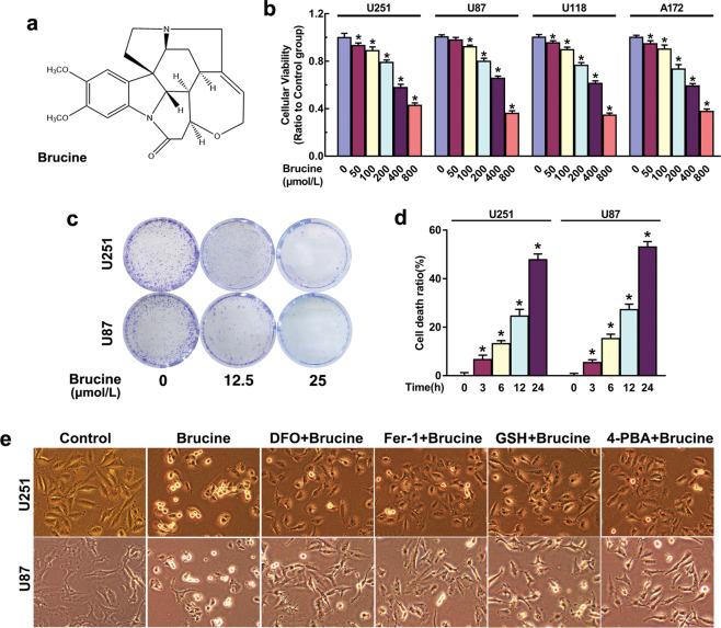
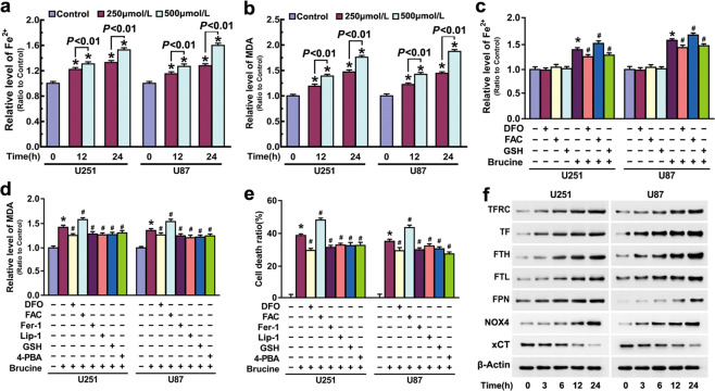
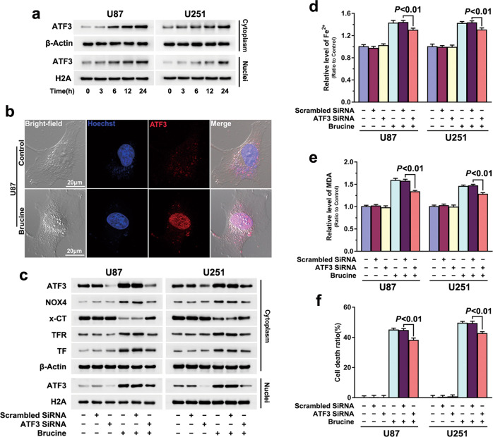
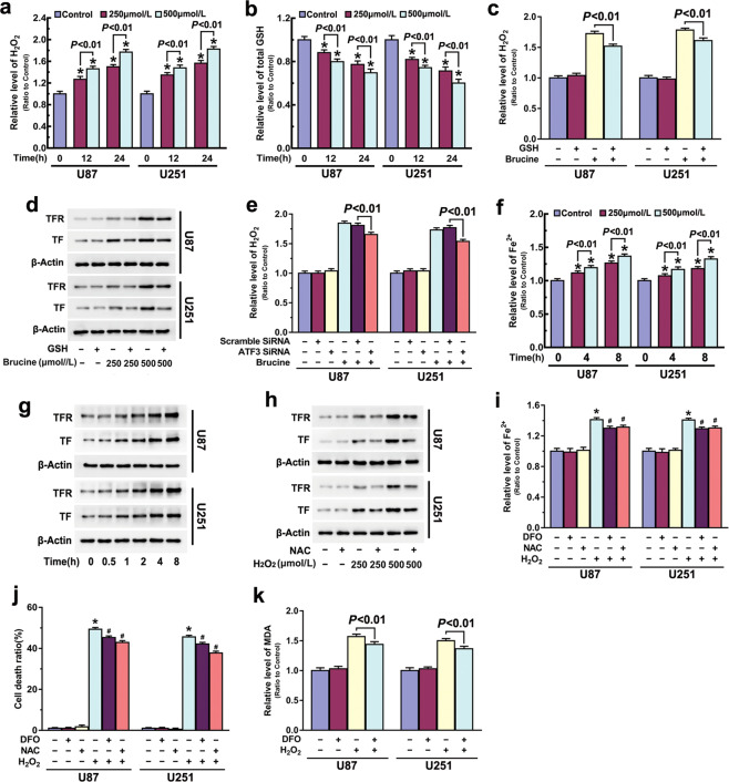
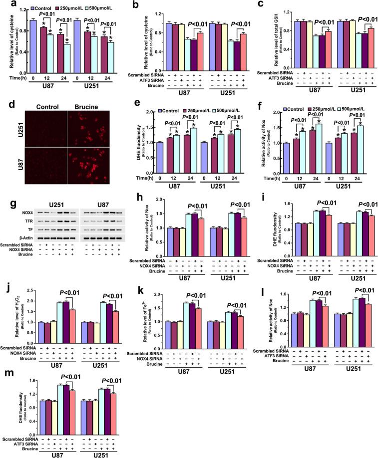
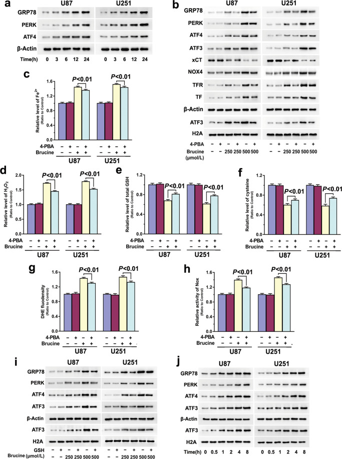
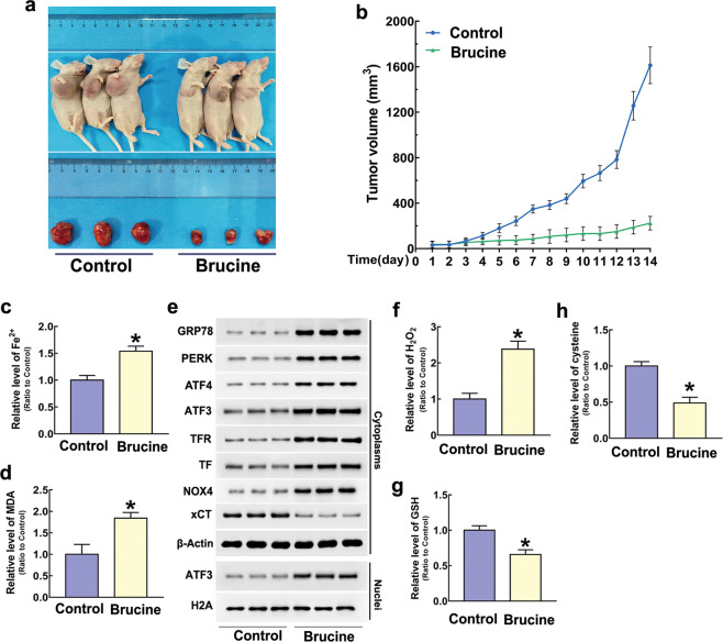
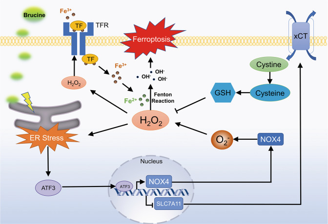
Ferroptotic cell death is characterized by iron-dependent lipid peroxidation that is initiated by ferrous iron and H2O2 via Fenton reaction, in which the role of activating transcription factor 3 (ATF3) remains elusive. Brucine is a weak alkaline indole alkaloid extracted from the seeds of Strychnos nux-vomica, which has shown potent antitumor activity against various tumors, including glioma. In this study, we showed that brucine inhibited glioma cell growth in vitro and in vivo, which was paralleled by nuclear translocation of ATF3, lipid peroxidation, and increases of iron and H2O2. Furthermore, brucine-induced lipid peroxidation was inhibited or exacerbated when intracellular iron was chelated by deferoxamine (500 μM) or improved by ferric ammonium citrate (500 μM). Suppression of lipid peroxidation with lipophilic antioxidants ferrostatin-1 (50 μM) or liproxstatin-1 (30 μM) rescued brucine-induced glioma cell death. Moreover, knockdown of ATF3 prevented brucine-induced accumulation of iron and H2O2 and glioma cell death. We revealed that brucine induced ATF3 upregulation and translocation into nuclei via activation of ER stress. ATF3 promoted brucine-induced H2O2 accumulation via upregulating NOX4 and SOD1 to generate H2O2 on one hand, and downregulating catalase and xCT to prevent H2O2 degradation on the other hand. H2O2 then contributed to brucine-triggered iron increase and transferrin receptor upregulation, as well as lipid peroxidation. This was further verified by treating glioma cells with exogenous H2O2 alone. Moreover, H2O2 reversely exacerbated brucine-induced ER stress. Taken together, ATF3 contributes to brucine-induced glioma cell ferroptosis via increasing H2O2 and iron.

Keywords: ATF3; ER stress; NOX4; brucine; ferroptosis; glioma; hydrogen peroxide.


Conflict of interest statement

The authors declare no competing interests.

Figures

Fig. 1
Fig. 1. Brucine inhibited glioma cell viability and induced glioma cell death.
a Chemical structure of brucine. b MTT showed that brucine inhibited the viabilities of human U251, U87, U118, and A172 glioma cells in a dosage-dependent manner. c Colony formation assay proved that 12.5 μM brucine could obviously inhibit U87 and U251 glioma cells to form colonies, which became more apparent when brucine dosage was increased to 25 μM. d LDH release assay showed that brucine at 500 μM induced glioma cell death in a time-dependent manner. e Representative images under microscope proved that the cells treated with brucine became shrunk and round when compared with control cells, which was apparently inhibited in the presence of deferoxamine (DFO), ferrostatin-1 (Fer-1), GSH, or 4-PBA. *P < 0.01 versus control group. The values are expressed as mean ± SEM (n = 5 per group).
Fig. 2
Fig. 2. Brucine induced ferroptosis in glioma cells.
a Iron assay showed that brucine improved intracellular ferrous iron in a time- and dosage-dependent manner. b MDA assay proved that brucine induced lipid peroxidation in a time- and dosage-dependent manner. c Iron assay revealed that brucine-induced increase of ferrous iron was inhibited by deferoxamine (DFO) and GSH, but reinforced by ferric ammonium citrate (FAC). d MDA assay demonstrated that brucine-induced lipid peroxidation was inhibited by deferoxamine (DFO), ferrostatin-1 (Fer-1), liproxstatin-1 (Lip-1), GSH, and 4-PBA, but aggravated by ferric ammonium citrate (FAC). e LDH release assay showed that brucine-induced glioma cell death was significantly inhibited by deferoxamine (DFO), ferrostatin-1 (Fer-1), liproxstatin-1 (Lip-1), GSH, and 4-PBA, but exacerbated by ferric ammonium citrate (FAC). f Western blotting analysis revealed that brucine triggered time-dependent upregulation of transferrin receptor (TFR), transferrin (TF), ferritin heavy chain (FTH), ferritin light chain (FTL), and NOX4 and downregulation of xCT. *P < 0.01 versus control group. #P < 0.01 versus brucine-treated group. The values are expressed as mean ± SEM (n = 5 per group).
Fig. 3
Fig. 3. ATF3 contributed to brucine-induced glioma cell ferroptosis.
a Western blotting analysis showed that brucine treatment resulted in time-dependent upregulation of ATF3 in both cytoplasmic and nuclear fractions in U87 and U251 glioma cells. b Representative images of confocal microscopy combined with immune-cytochemistry staining confirmed that ATF3 accumulated apparently in nucleus of the U87 cell treated with brucine for 24 h. c Western blotting revealed that knockdown of ATF3 with siRNA prevented brucine-triggered upregulation of NOX4, transferrin, and transferrin receptor and downregulation of xCT. d Iron assay proved that ATF3 knockdown prevented iron increase caused by brucine. e MDA assay showed that ATF3 knockdown inhibited brucine-induced lipid peroxidation. f LDH release assay demonstrated that brucine-induced glioma cell death was significantly prevented when ATF3 was knocked down with siRNA. The values are expressed as mean ± SEM (n = 5 per group)
Fig. 4
Fig. 4. ATF3 contributed to brucine-induced H2O2 increase.
a Brucine induced accumulation of H2O2 in a time- and dosage-dependent manner. b Brucine treatment resulted in depletion of GSH in a time- and dosage-dependent manner. c Supplement of GSH prevented brucine-induced accumulation of H2O2. d Western blotting revealed that brucine-induced increases of transferrin (TF) and transferrin receptor (TFR) were both inhibited when GSH was supplemented. e Knockdown of ATF3 with siRNA prevented brucine-induced accumulation of H2O2. f Iron assay showed that ferrous iron was improved in the cells treated with H2O2 alone in a time- and dosage-dependent manner. g Western blotting revealed that H2O2 induced time-dependent upregulation of transferrin receptor and transferrin in U87 and U251 glioma cells. h Western blotting showed that H2O2-induced upregulation of transferrin receptor and transferrin was obviously inhibited in the presence of antioxidant NAC. i Iron assay showed that H2O2 induced apparent improvement of ferrous iron, which was inhibited in the cells pretreated with deferoxamine (DFO) and NAC. j LDH release assay demonstrated that H2O2-induced glioma cell death was apparently inhibited by deferoxamine (DFO) or NAC. k MDA assay demonstrated that H2O2 alone could induce lipid peroxidation, which was suppressed in the presence of deferoxamine (DFO). *P < 0.01 versus control group. The values are expressed as mean ± SEM (n = 5 per group).
Fig. 5
Fig. 5. ATF3 regulated brucine-induced NOX4 upregulation and xCT downregulation.
a Brucine induced time- and dosage-dependent depletion of cysteine in glioma cells. b Knockdown of ATF3 with siRNA prevented brucine-induced depletion of cysteine. c ATF3 knockdown inhibited brucine-induced GSH depletion. d Representative images of fluorescence microscopy showed that the red fluorescence exhibited by DHE was much stronger in brucine-treated cells when compared with that in control cells. e Statistical analysis of the fluorescence intensity proved that brucine improved superoxide in a time- and dosage-dependent manner. f NADPH oxidase activity assay showed that brucine activated NADPH oxidase in a time- and dosage-dependent manner. g Western blotting analysis proved that knockdown of NOX4 with siRNA prevented brucine-triggered upregulation of transferrin (TF) and transferrin receptor (TFR). h Brucine-induced upregulation in the activities of NADPH oxidases was suppressed when NOX4 was knocked down. i Knockdown of NOX4 prevented brucine-triggered increase of superoxide. j Brucine-induced accumulation of H2O2 was inhibited when NOX4 was knocked down. k Knockdown of NOX4 prevented the increase of ferrous iron caused by brucine. l Brucine-induced upregulation in NADPH oxidases activities was suppressed when ATF3 was knocked down with siRNA. m Knockdown of ATF3 prevented brucine-induced increase of superoxide. The values are expressed as mean ± SEM (n = 5 per group).
Fig. 6
Fig. 6. Brucine induced a positive feedback between ER stress and H2O2 generation.
a Western blotting showed that brucine upregulated GRP78, PERK, and ATF4 in glioma cells in a time-dependent manner. b Western blotting revealed that pretreatment with 4-PBA obviously inhibited brucine-induced upregulation of GRP78, PERK, ATF4, NOX4, transferrin (TF) and transferrin receptor (TFR), downregulation of xCT, and nuclear translocation of ATF3. c Iron assay demonstrated that brucine-induced increase of ferrous iron was significantly inhibited by 4-PBA. d 4-PBA prevented brucine-induced accumulation of H2O2. e Brucine-induced GSH depletion was suppressed in the presence of 4-PBA. f The depletion of cysteine triggered by brucine was prevented in the cells pretreated with 4-PBA. g Brucine-induced improvement of superoxide was alleviated by pretreatment with 4-PBA. h 4-PBA obviously inhibited brucine-induced activation of NADPH oxidases. i Western blotting revealed that 4-PBA prevented brucine-induced upregulation of GRP78, PERK, and ATF4, as well as nuclear translocation of ATF3. j H2O2 could induce upregulation of GRP78, PERK, and ATF4 and nuclear translocation of ATF3. The values are expressed as mean ± SEM (n = 5 per group).
Fig. 7
Fig. 7. Brucine induced increases of iron and H2O2 and lipid peroxidation in vivo.
a Representative images of the nude mice with xenografted gliomas showed that tumor size was significantly smaller in the mice treated with brucine at the dosage of 40 mg/kg for consecutive 13 days than that in control group. b Statistical analysis of tumor volumes confirmed as well that TMZ inhibited tumor growth in vivo. c Iron assay showed ferrous iron level was significantly higher in brucine-treated group than that in control group. d MDA assay proved that lipid peroxidation became more apparent in brucine-treated group when compared with control group. e Western blotting analysis revealed that brucine induced marked upregulation of GRP78, PERK, ATF4, NOX4, transferrin (TF), and transferrin receptor (TFR) and downregulation of xCT and nuclear translocation of ATF3. f The level of H2O2 was increased obviously by brucine in vivo. g GSH was decreased significantly in brucine-treated group when compared with control group. h Cysteine was depleted by cysteine in vivo. *P < 0.01 versus control group. The values are expressed as mean ± SEM (n = 6 per group).
Fig. 8
Fig. 8. Schematic diagram for the role of ATF3 in brucine-induced glioma cell ferroptosis.
Brucine treatment promoted ATF3 upregulation and translocation into nucleus via causing ER stress. Then, ATF3 improved intracellular level of H2O2 via two pathways. One is to activate NOX4 transcription to excessively generate superoxide, and the other is to repress the transcription of SLC7A11 to deplete cysteine and GSH by inhibition of xCT. The improved H2O2 not only increased intracellular iron by upregulation of the expression of transferrin receptor, but also reacted with iron via fenton reaction to generate toxic hydroxyl radicals. Eventually, hydroxyl radicals contributed to glioma cell death by causing lipid peroxidation.

References

    1. Lapointe S, Perry A, Butowski NA. Primary brain tumours in adults. Lancet. 2018;392:432–46.. - PubMed
    1. Messaoudi K, Clavreul A, Lagarce F. Toward an effective strategy in glioblastoma treatment. Part I: resistance mechanisms and strategies to overcome resistance of glioblastoma to temozolomide. Drug Discov Today. 2015;20:899–905. doi: 10.1016/j.drudis.2015.02.011. - DOI - PubMed
    1. Dixon SJ, Lemberg KM, Lamprecht MR, Skouta R, Zaitsev EM, Gleason CE, et al. Ferroptosis: an iron-dependent form of nonapoptotic cell death. Cell. 2012;149:1060–72. doi: 10.1016/j.cell.2012.03.042. - DOI - PMC - PubMed
    1. Chen X, Li J, Kang R, Klionsky DJ, Tang D. Ferroptosis: machinery and regulation. Autophagy. 2020;27:1–28. 10.1080/15548627.2020.1810918. - PMC - PubMed
    1. Torti SV, Torti FM. Iron and cancer: more ore to be mined. Nat Rev Cancer. 2013;13:342–55. doi: 10.1038/nrc3495. - DOI - PMC - PubMed

MeSH terms