. 2021 Oct;23(10):1912-1921.
doi: 10.1038/s41436-021-01222-w. Epub 2021 Jun 10.

Truncating variants in the SHANK1 gene are associated with a spectrum of neurodevelopmental disorders

Affiliations

Truncating variants in the SHANK1 gene are associated with a spectrum of neurodevelopmental disorders

Halie J May et al. Genet Med. 2021 Oct.

Abstract

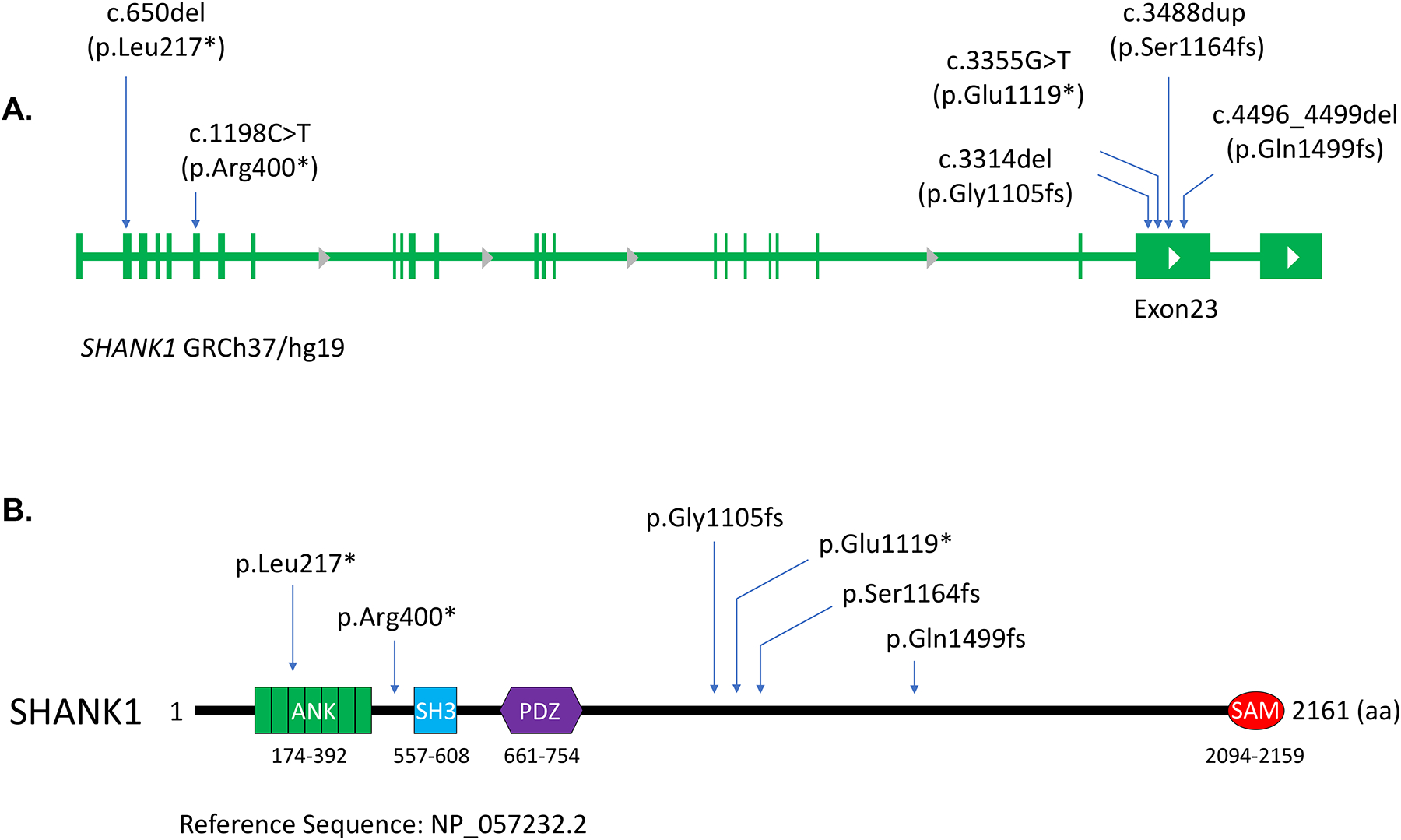
Purpose: In this study, we aimed to characterize the clinical phenotype of a SHANK1-related disorder and define the functional consequences of SHANK1 truncating variants.

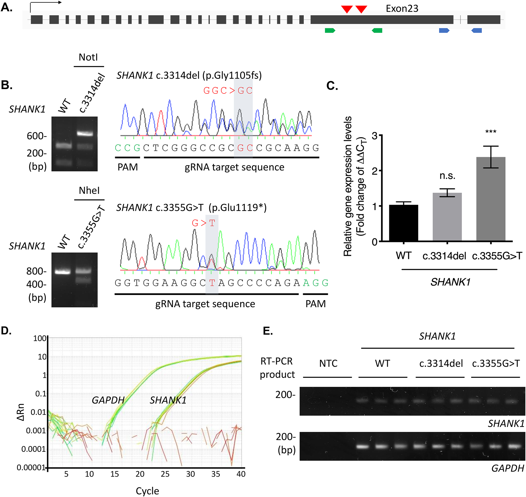
Methods: Exome sequencing (ES) was performed for six individuals who presented with neurodevelopmental disorders. Individuals were ascertained with the use of GeneMatcher and Database of Chromosomal Imbalance and Phenotype in Humans Using Ensembl Resources (DECIPHER). We evaluated potential nonsense-mediated decay (NMD) of two variants by making knock-in cell lines of endogenous truncated SHANK1, and expressed the truncated SHANK1 complementary DNA (cDNA) in HEK293 cells and cultured hippocampal neurons to examine the proteins.

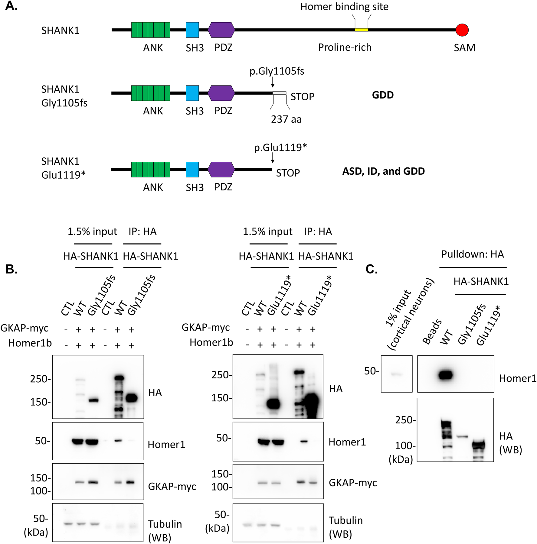
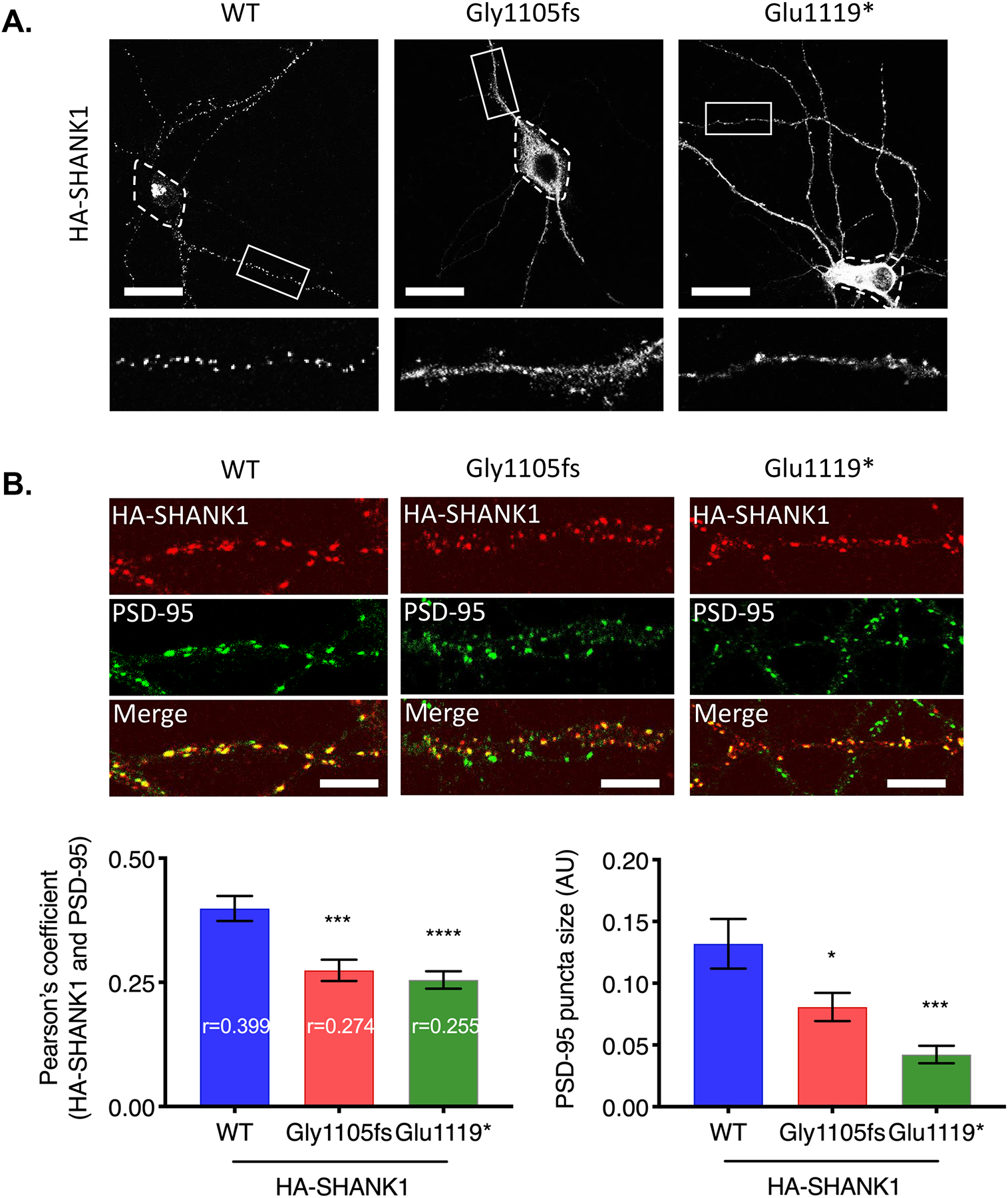
Results: ES detected de novo truncating variants in SHANK1 in six individuals. Evaluation of NMD resulted in stable transcripts, and the truncated SHANK1 completely lost binding with Homer1, a linker protein that binds to the C-terminus of SHANK1. These variants may disrupt protein-protein networks in dendritic spines. Dispersed localization of the truncated SHANK1 variants within the spine and dendritic shaft was also observed when expressed in neurons, indicating impaired synaptic localization of truncated SHANK1.

Conclusion: This report expands the clinical spectrum of individuals with truncating SHANK1 variants and describes the impact these variants may have on the pathophysiology of neurodevelopmental disorders.


Conflict of interest statement

Conflicts of Interest

Aida Telegrafi and Richard Person are employees of GeneDx, Inc. David Goldstein is a founder of and holds equity in Q State Biosciences and Praxis Therapeutics; holds equity in Apostle Inc., and serves as a consultant to AstraZeneca, Gilead Sciences, GoldFinch Bio and Gossamer Bio. No other authors have conflicting interests to disclose.

Figures

Figure 1.
Figure 1.. De novo SHANK1 variants identified in patients with developmental delay and autism.
(A) Schematic figures show the distribution of six de novo variants observed in our cohort in the SHANK1 genome. (B) Amino acid changes caused by the SHANK1 variants are indicated in the diagram of the SHANK1 protein. Protein domains and amino acid numbers are indicated.
Figure 2.
Figure 2.. SHANK1 truncating variants were transcribed in CRISPR KI HEK293 cell lines.
(A) Two de novo SHANK1 variants, c.3314del (p.Gly1105fs) and c.3355G>T (p.Glu1119*), in the penultimate exon of SHANK1 gene are selected for generation of CRISPR KI cell lines. Red arrow heads indicate brief location of the variants in the SHANK1 genome. Green arrows indicate binding sites of the primer set for genotyping. Blue arrows represent exons spanning primers binding sites for real-time PCR. (B) gDNA prepared from the SHANK1 c.3314del CRISPR and c.3355G>T CRISPR cell lines were amplified for the KI region by PCR. The PCR products were digested by NotI restriction enzyme and sequenced to check SHANK1 c.3314delG (top). The PCR products were digested with NheI restriction enzyme and sequenced to confirm SHANK1 c.3355G>T (bottom). (C) Total transcripts were prepared from the CRISPR cell lines and real-time PCR was performed. Fold change of the SHANK1 transcript levels (2^-(ΔΔCT)) was calculated. Graph indicates mean ± SEM (n = 9). The statistical significance between the mean of WT and the mean of each SHANK1 variant was calculated using one-way ANOVA with Dunnett’s multiple comparison test. ***P = 0.0001. n.s., not significant. (D) Representative SYBR green amplification graphs of real-time PCR analysis for SHANK1 and GAPDH. (E) Final product of the real-time PCR were run onto 2% agarose gel to check specific amplification.
Figure 3.
Figure 3.. Truncated SHANK1 proteins abolished Homer1 binding.
(A) Schematic figures denote SHANK1 c.3314del (p.Gly1105fs) and c.3355G>T (p.Glu1119*) variants and resulting SHANK1 protein truncations. (B) HA-SHANK1 (WT, Gly1105fs, or Glu1119*), GKAP-myc and Homer1b were co-transfected in HEK293 cells as indicated in the figures. GKAP-myc and Homer1b in the immunoprecipitates with HA antibody were analyzed by immunoblotting. (C) The immunoprecipitated HA-SHANK1 (WT, Gly1105fs, or Glu1119*) beads from HEK293 cells were incubated with cultured rat cortical neuron lysates for pulldown assays. Endogenous Homer1 binding with the immunoprecipitated HA-SHANK1 was analyzed by immunoblotting.
Figure 4.
Figure 4.. Truncated SHANK1 proteins disrupted synaptic localization.
(A) HA-SHANK1 (WT, Gly1105fs, or Glu1119*) was transfected in cultured rat hippocampal neurons. HA-SHANK1 was labeled with anti-HA and Alexa 555-conjugated secondary antibody (white). Enlarged images of the boxed regions are shown below each panel. Dashed regions indicate soma. (Scale bar, 25 μm.) (B) HA-SHANK1 (WT, Gly1105fs, or Glu1119*) was expressed in cultured rat hippocampal neurons. HA-SHANK1 was labeled with anti-HA and Alexa 555-conjugated secondary antibody (red). Endogenous PSD-95 was labeled with anti-PSD-95 and Alexa 488-conjugated secondary antibody (green). Regions from three dendrites per each neuron were analyzed for Pearson’s coefficient. Graph indicates mean ± SEM (n = 15~19). *P < 0.05, ***P < 0.0005, ****P < 0.0001 using one-way ANOVA with Dunnett’s multiple comparison test. (Scale bar, 5 μm.)

References

    1. Sheng M, Kim E. The Shank family of scaffold proteins. J Cell Sci. 2000;113 (Pt 11):1851–1856. - PubMed
    1. Mao W, Watanabe T, Cho S, et al.Shank1 regulates excitatory synaptic transmission in mouse hippocampal parvalbumin-expressing inhibitory interneurons. Eur J Neurosci. 2015;41(8):1025–1035. - PMC - PubMed
    1. Romorini S, Piccoli G, Jiang M, et al.A functional role of postsynaptic density-95-guanylate kinase-associated protein complex in regulating Shank assembly and stability to synapses. J Neurosci. 2004;24(42):9391–9404. - PMC - PubMed
    1. Sala C, Piech V, Wilson NR, Passafaro M, Liu G, Sheng M. Regulation of dendritic spine morphology and synaptic function by Shank and Homer. Neuron. 2001;31(1):115–130. - PubMed
    1. Hung AY, Futai K, Sala C, et al.Smaller dendritic spines, weaker synaptic transmission, but enhanced spatial learning in mice lacking Shank1. J Neurosci. 2008;28(7):1697–1708. - PMC - PubMed

Web Resources

    1. ClinVar browser: http://www.ncbi.nlm.nih.gov/clinvar/
    1. Consensus Coding Sequence (CCDS), https://www.ncbi.nlm.nih.gov/CCDS/
    1. Ensembl genome assembly GRCh37: http://grch37.ensembl.org/Homo_sapiens/Info/Index.
    1. Ensembl Variant Effect Predictor (VEP): http://grch37.ensembl.org/Homo_sapiens/Tools/VEP.
    1. GenBank, https://www.ncbi.nlm.nih.gov/genbank/

Publication types

Substances