Review
. 2021 May 25:15:658202.
doi: 10.3389/fnins.2021.658202. eCollection 2021.

Kynurenines and Neurofilament Light Chain in Multiple Sclerosis

Affiliations
Review

Kynurenines and Neurofilament Light Chain in Multiple Sclerosis

Dániel Pukoli et al. Front Neurosci. .

Abstract

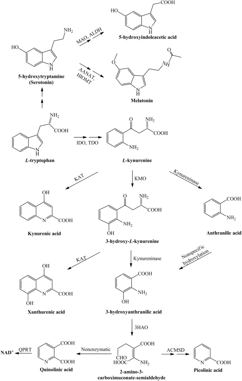
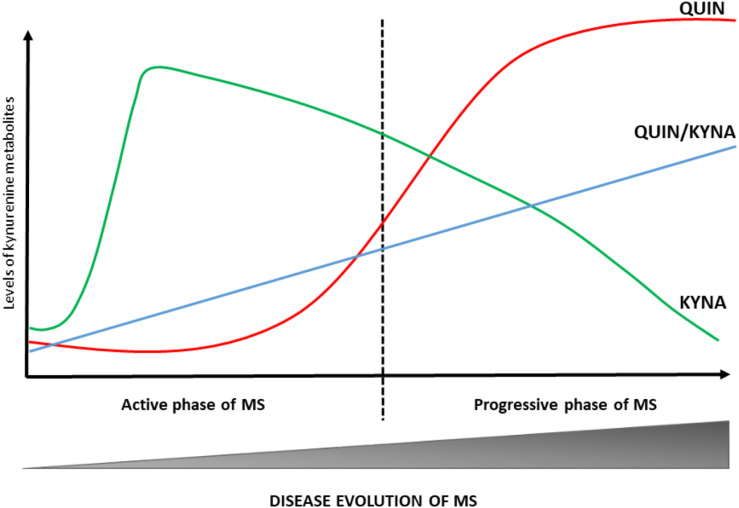
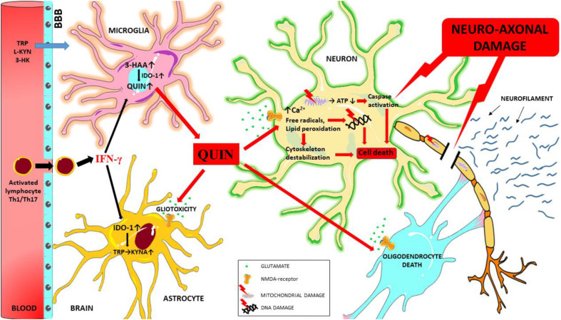
Multiple sclerosis is an autoimmune, demyelinating, and neurodegenerative disease of the central nervous system. In recent years, it has been proven that the kynurenine system plays a significant role in the development of several nervous system disorders, including multiple sclerosis. Kynurenine pathway metabolites have both neurotoxic and neuroprotective effects. Moreover, the enzymes of the kynurenine pathway play an important role in immunomodulation processes, among others, as well as interacting with neuronal energy balance and various redox reactions. Dysregulation of many of the enzymatic steps in kynurenine pathway and upregulated levels of these metabolites locally in the central nervous system, contribute to the progression of multiple sclerosis pathology. This process can initiate a pathogenic cascade, including microglia activation, glutamate excitotoxicity, chronic oxidative stress or accumulated mitochondrial damage in the axons, that finally disrupt the homeostasis of neurons, leads to destabilization of neuronal cell cytoskeleton, contributes to neuro-axonal damage and neurodegeneration. Neurofilaments are good biomarkers of the neuro-axonal damage and their level reliably indicates the severity of multiple sclerosis and the treatment response. There is increasing evidence that connections exist between the molecules generated in the kynurenine metabolic pathway and the change in neurofilament concentrations. Thus the alterations in the kynurenine pathway may be an important biomarker of the course of multiple sclerosis. In our present review, we report the possible relationship and connection between neurofilaments and the kynurenine system in multiple sclerosis based on the available evidences.

Keywords: glutamate excitotoxicity; kynurenine; mitochondrial dysfunction; multiple sclerosis; neuro-axonal damage; neurofilaments; oxidative stress; quinolinic acid.


Conflict of interest statement

The authors declare that the research was conducted in the absence of any commercial or financial relationships that could be construed as a potential conflict of interest.

Figures

FIGURE 1
FIGURE 1
Detailed representation of the kynurenine pathway. Tryptophan, as an essential amino acid, is a precursor of the serotonin- and kynurenine pathway. During the serotonin pathway, melatonin and 5-hydroxyindoleacetic acid are formed, while the kynurenine pathway of tryptophan metabolism leads to the production of nicotinamide adenine dinucleotide. The conversion of tryptophan to L-kynurenine is performed by IDO/TDO. From L-kynurenine various neuroactive metabolites are formed during this pathway, which eventually leads to the nicotinamide adenine dinucleotide, that plays a significant role in the production of cellular energy. 3-HAO, 3-hydroxyanthranilate oxidase; AANAT, arylalkylamine N-acetyltransferase; ACMSD, α-amino-β-carboxymuconate-semialdehyde-decarboxylase; ALDH, aldehyde dehydrogenase; HIOMT, hydroxyindole-O-methyltransferase; IDO, indoleamine 2,3-dioxygenase; KAT, kynurenine aminotransferase; KMO, kynurenine 3-monooxygenase; MAO, monoamine oxidase; NAD+, nicotinamide adenine dinucleotide; QPRT, quinolinate phosphoribosyltransferase; TDO, tryptophan 2,3-dioxygenase.
FIGURE 2
FIGURE 2
Changes in KP profile during disease course of MS. The KP metabolic profile changes at different stages of MS. In the active phase of MS, the KYNA levels are increased. During the progressive phase of the disease QUIN levels and QUIN/KYNA ratio are increased. These changes in metabolic profile of the KP have a strong association with disease activity. KYNA, kynurenic acid; MS, multiple sclerosis; QUIN, quinolinic acid.
FIGURE 3
FIGURE 3
Schematic overview of the mechanisms of QUIN toxicity in MS. During neuroinflammation autoreactive T cells (Th1 and Th17) and activated monocytes/macrophages migrate to the central nervous system through the damaged blood-brain barrier, and these cells secrete numerous pro-inflammatory cytokines (such as interferon gamma). Interferon gamma is one of the most potent activators of IDO-1, the primary enzyme of the KP. In case of an enzymatic imbalance in the KP and also through the chronic alteration of the physiological concentration of kynurenine metabolites in the microenvironment the balance between neuroprotective and neurotoxic processes is no longer ensured. Neurotoxic QUIN secreted in abnormal quantities by activated macrophages and microglia cells in an inflammatory microenvironment can damage oligodendrocytes, astrocytes and neuron through multiple mechanisms. QUIN induces caspase activation, mitochondrial impairment, oxidative stress, lipid peroxidation and energy deficit through the overactivation of NMDA receptors (and also independently of overactivation). These factors combine to result in the destabilization of the cytoskeleton, leading to DNA damage, cell death and ultimately neuro-axonal damage. Neurofilaments are released as a result of neuro-axonal injury. 3-HAA, 3-hydroxyantranilic acid; 3-HK, 3-hydroxykynurenine; ATP, adenosine triphosphate; BBB, blood-brain barrier; IFN-γ, interferon gamma; IDO-1, indoleamine 2,3-dioxygenase; L-KYN, L-kynurenine; KYNA, kynurenic acid; QUIN, quinolinic acid; TRP, tryptophan.

References

    1. Aeinehband S., Brenner P., Ståhl S., Bhat M., Fidock M. D., Khademi M., et al. (2016). Cerebrospinal fluid kynurenines in multiple sclerosis; relation to disease course and neurocognitive symptoms. Brain Behav. Immun. 51 47–55. 10.1016/j.bbi.2015.07.016 - DOI - PubMed
    1. Amirkhani A., Rajda C., Arvidsson B., Bencsik K., Boda K., Seres E., et al. (2005). Interferon-beta affects the tryptophan metabolism in multiple sclerosis patients. Eur. J. Neurol. 12 625–631. 10.1111/j.1468-1331.2005.01041.x - DOI - PubMed
    1. Anderson J. M., Hampton D. W., Patani R., Pryce G., Crowther R. A., Reynolds R., et al. (2008). Abnormally phosphorylated tau is associated with neuronal and axonal loss in experimental autoimmune encephalomyelitis and multiple sclerosis. Brain 131 (Pt 7) 1736–1748. 10.1093/brain/awn119 - DOI - PubMed
    1. Aranami T., Yamamura T. (2008). Th17 Cells and autoimmune encephalomyelitis (EAE/MS). Allergol. Int. 57 115–120. 10.2332/allergolint.R-07-159 - DOI - PubMed
    1. Barro C., Benkert P., Disanto G., Tsagkas C., Amann M., Naegelin Y., et al. (2018). Serum neurofilament as a predictor of disease worsening and brain and spinal cord atrophy in multiple sclerosis. Brain 141 2382–2391. 10.1093/brain/awy154 - DOI - PubMed

LinkOut - more resources