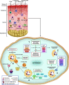
Autophagy as a Target for Drug Development Of Skin Infection Caused by Mycobacteria
- PMID: 34113346
- PMCID: PMC8185338
- DOI: 10.3389/fimmu.2021.674241
Autophagy as a Target for Drug Development Of Skin Infection Caused by Mycobacteria
Abstract
Pathogenic mycobacteria species may subvert the innate immune mechanisms and can modulate the activation of cells that cause disease in the skin. Cutaneous mycobacterial infection may present different clinical presentations and it is associated with stigma, deformity, and disability. The understanding of the immunopathogenic mechanisms related to mycobacterial infection in human skin is of pivotal importance to identify targets for new therapeutic strategies. The occurrence of reactional episodes and relapse in leprosy patients, the emergence of resistant mycobacteria strains, and the absence of effective drugs to treat mycobacterial cutaneous infection increased the interest in the development of therapies based on repurposed drugs against mycobacteria. The mechanism of action of many of these therapies evaluated is linked to the activation of autophagy. Autophagy is an evolutionary conserved lysosomal degradation pathway that has been associated with the control of the mycobacterial bacillary load. Here, we review the role of autophagy in the pathogenesis of cutaneous mycobacterial infection and discuss the perspectives of autophagy as a target for drug development and repurposing against cutaneous mycobacterial infection.
Keywords: autophagy; drug development; mycobacteria; skin; skin cells.
Copyright © 2021 Bittencourt, da Silva Prata, de Andrade Silva, de Mattos Barbosa, Dalcolmo and Pinheiro.
Conflict of interest statement
The authors declare that the research was conducted in the absence of any commercial or financial relationships that could be construed as a potential conflict of interest.
Figures

Similar articles
-
Cutaneous Mycobacterial Infections.Clin Microbiol Rev. 2018 Nov 14;32(1):e00069-18. doi: 10.1128/CMR.00069-18. Print 2018 Jan. Clin Microbiol Rev. 2018. PMID: 30429139 Free PMC article. Review.
-
Cutaneous infection with rapidly-growing mycobacterial infection following heart transplant: a case report and review of the literature.Transplant Proc. 2006 Jun;38(5):1526-9. doi: 10.1016/j.transproceed.2006.02.126. Transplant Proc. 2006. PMID: 16797350 Review.
-
Nontuberculous mycobacterial infections of the skin: A retrospective study of 25 cases.J Am Acad Dermatol. 2007 Sep;57(3):413-20. doi: 10.1016/j.jaad.2007.01.042. Epub 2007 Mar 26. J Am Acad Dermatol. 2007. PMID: 17368631
-
Cutaneous non-tuberculous Mycobacterial infections: a clinical and histopathological study of 17 cases from Lebanon.J Eur Acad Dermatol Venereol. 2011 Jan;25(1):33-42. doi: 10.1111/j.1468-3083.2010.03684.x. J Eur Acad Dermatol Venereol. 2011. PMID: 20456544
-
Cutaneous infection due to Mycobacterium immunogenum: an European case report and review of the literature.Dermatol Online J. 2017 Oct 15;23(10):13030/qt9zg5r07t. Dermatol Online J. 2017. PMID: 29469779 Review.
Cited by
-
Targeting Autophagy as a Strategy for Developing New Host-Directed Therapeutics Against Nontuberculous Mycobacteria.Pathogens. 2025 May 13;14(5):472. doi: 10.3390/pathogens14050472. Pathogens. 2025. PMID: 40430792 Free PMC article. Review.
-
Potential Role of CXCL10 in Monitoring Response to Treatment in Leprosy Patients.Front Immunol. 2021 Jul 20;12:662307. doi: 10.3389/fimmu.2021.662307. eCollection 2021. Front Immunol. 2021. PMID: 34354699 Free PMC article.
-
The role of Bcl‑2 in controlling the transition between autophagy and apoptosis (Review).Mol Med Rep. 2025 Jul;32(1):172. doi: 10.3892/mmr.2025.13537. Epub 2025 Apr 17. Mol Med Rep. 2025. PMID: 40242969 Free PMC article. Review.
-
Cell Death Mechanisms in Mycobacterium abscessus Infection: A Double-Edged Sword.Pathogens. 2025 Apr 16;14(4):391. doi: 10.3390/pathogens14040391. Pathogens. 2025. PMID: 40333197 Free PMC article. Review.
-
Investigating cutaneous tuberculosis and nontuberculous mycobacterial infections a Department of Dermatology, Beijing, China: a comprehensive clinicopathological analysis.Front Cell Infect Microbiol. 2024 Aug 23;14:1451602. doi: 10.3389/fcimb.2024.1451602. eCollection 2024. Front Cell Infect Microbiol. 2024. PMID: 39247053 Free PMC article.
References
Publication types
MeSH terms
LinkOut - more resources
Full Text Sources
Medical

