

Antitumor Effects of Freeze-Dried Robusta Coffee (Coffea canephora) Extracts on Breast Cancer Cell Lines
- PMID: 34113419
- PMCID: PMC8154281
- DOI: 10.1155/2021/5572630
Antitumor Effects of Freeze-Dried Robusta Coffee (Coffea canephora) Extracts on Breast Cancer Cell Lines
Abstract
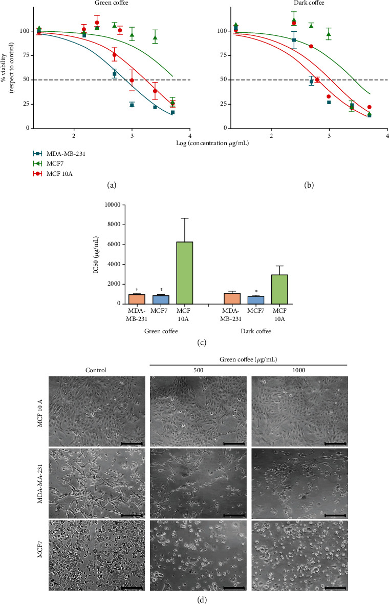
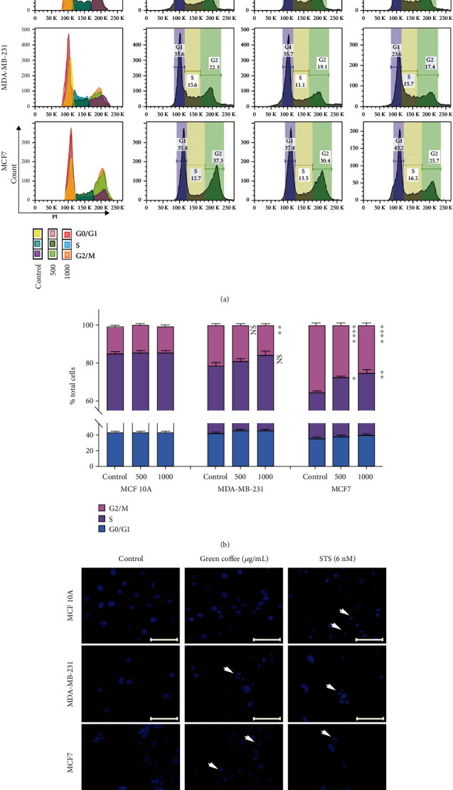
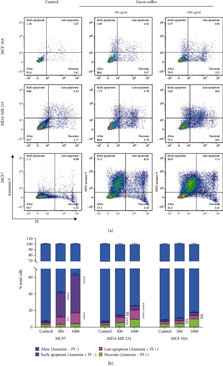
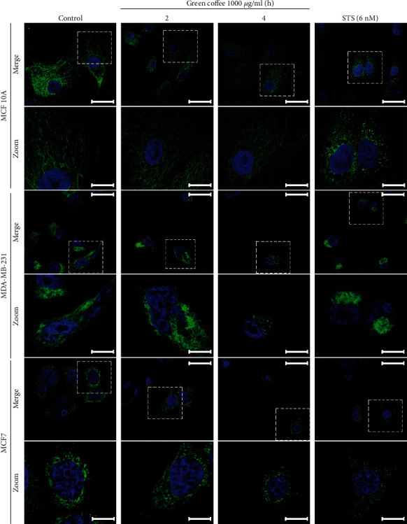
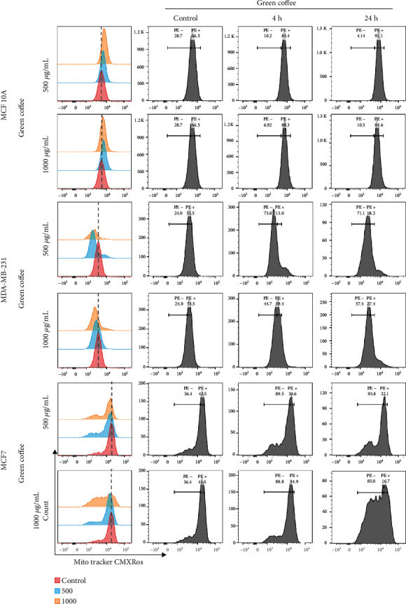
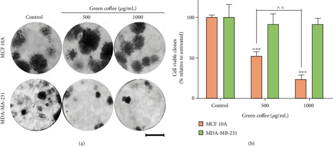
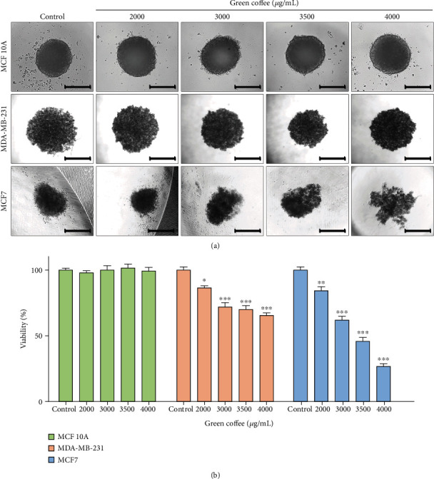
Coffee consumption is believed to have chemopreventive and chemotherapeutic effects and to contribute to preventing the development and progression of cancer. However, there is still controversy around these claims. As indicated in our previous works, diet can influence the risk of breast cancer. Intake of coffee is hypothesized to reduce this risk, but current scientific evidence is not conclusive. This work is aimed at studying the effects of Robusta coffee bean extract on cell viability, proliferation, and apoptosis of different human cancers, especially breast cancer cell lines. To this end, cell viability was evaluated by Alamar Blue in 2D and 3D models, the cell cycle by PI, apoptosis by annexin V, mitochondrial morphology, and functionality by mitoTracker, and colony formation capacity by the clonogenic assay. Green and dark coffee extract significantly reduced viability in human breast, colorectal, brain, and bone cancer cells. Coffee anticancer activity was clearly evidenced in MDA-MB-231 (ER-) and MCF-7 (ER+) breast cancer cells but not in the normal breast cell line. In addition, coffee extract induces an increase S phase and a decrease G2/M population in breast cancer cells, affected the mitochondrial morphology, and triggered apoptosis. MDA-MB-231 breast cancer cells lost their clonogenic capacity after treatment. The antitumor activity was demonstrated in both 2D and 3D culture cell models.
Copyright © 2021 Ayelén D. Nigra et al.
Conflict of interest statement
The authors declare that they have no known competing financial interests or personal relationships that could influence the work reported in this paper.
Figures
Similar articles
-
Effect of Roasting Levels and Drying Process of Coffea canephora on the Quality of Bioactive Compounds and Cytotoxicity.Int J Mol Sci. 2018 Oct 31;19(11):3407. doi: 10.3390/ijms19113407. Int J Mol Sci. 2018. PMID: 30384410 Free PMC article.
-
Immunoexpression of aortic endothelial P-selectin and serum apolipoprotein A-1 levels after administration of arabica (Coffea arabica) and robusta (Coffea canephora) coffee bean extracts: In vivo study in atherosclerosis rat model.Narra J. 2024 Aug;4(2):e794. doi: 10.52225/narra.v4i2.794. Epub 2024 Aug 2. Narra J. 2024. PMID: 39280317 Free PMC article.
-
Quantification of Coffea arabica and Coffea canephora var. robusta in roasted and ground coffee blends.Talanta. 2013 Mar 15;106:169-73. doi: 10.1016/j.talanta.2012.12.003. Epub 2012 Dec 23. Talanta. 2013. PMID: 23598112
-
Rapid authentication of coffee blends and quantification of 16-O-methylcafestol in roasted coffee beans by nuclear magnetic resonance.J Agric Food Chem. 2014 Dec 24;62(51):12309-14. doi: 10.1021/jf505013d. Epub 2014 Dec 10. J Agric Food Chem. 2014. PMID: 25431971
-
Furan in roasted, ground and brewed coffee.Rocz Panstw Zakl Hig. 2018;69(2):111-118. Rocz Panstw Zakl Hig. 2018. PMID: 29766689 Review.
Cited by
-
A Decade of Research on Coffee as an Anticarcinogenic Beverage.Oxid Med Cell Longev. 2021 Sep 15;2021:4420479. doi: 10.1155/2021/4420479. eCollection 2021. Oxid Med Cell Longev. 2021. PMID: 34567408 Free PMC article. Review.
-
Self-reported and genetically predicted effects of coffee intake on rheumatoid arthritis: Epidemiological studies and Mendelian randomization analysis.Front Nutr. 2022 Sep 12;9:926190. doi: 10.3389/fnut.2022.926190. eCollection 2022. Front Nutr. 2022. PMID: 36172525 Free PMC article.
-
Phytochemistry, Anti-cancer, and Anti-diabetic Properties of Plant-Based Foods from Mexican Agrobiodiversity: A Review.Foods. 2024 Dec 23;13(24):4176. doi: 10.3390/foods13244176. Foods. 2024. PMID: 39767118 Free PMC article. Review.
-
Turkish coffee has an antitumor effect on breast cancer cells in vitro and in vivo.Nutr Metab (Lond). 2024 Sep 13;21(1):73. doi: 10.1186/s12986-024-00846-4. Nutr Metab (Lond). 2024. PMID: 39272080 Free PMC article.
-
Onco-Breastomics: An Eco-Evo-Devo Holistic Approach.Int J Mol Sci. 2024 Jan 28;25(3):1628. doi: 10.3390/ijms25031628. Int J Mol Sci. 2024. PMID: 38338903 Free PMC article. Review.
References
MeSH terms
Substances
LinkOut - more resources
Full Text Sources
Medical
Research Materials
Miscellaneous
