

Membrane protein mediated bilayer communication in networks of droplet interface bilayers
- PMID: 34113722
- PMCID: PMC7610947
- DOI: 10.1038/s42004-020-0322-1
Membrane protein mediated bilayer communication in networks of droplet interface bilayers
Abstract
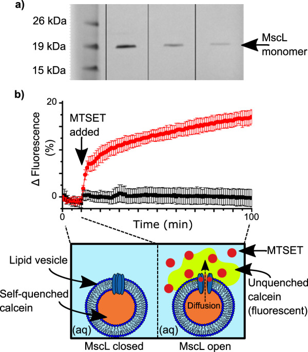
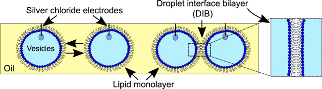
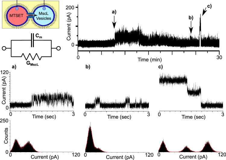
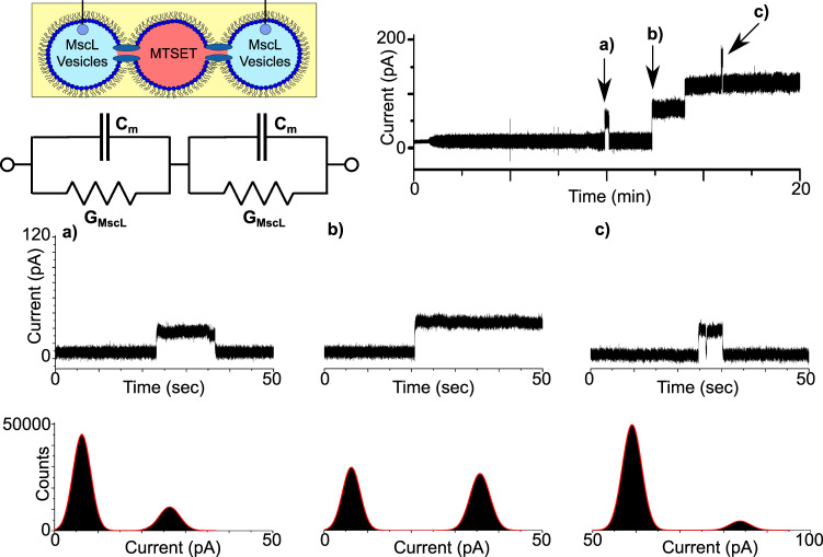
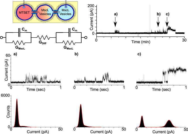
Droplet interface bilayers (DIBs) are model membranes formed between lipid monolayer-encased water droplets in oil. Compared to conventional methods, one of the most unique properties of DIBs is that they can be connected together to generate multi-layered 'tissue-like' networks, however introducing communication pathways between these compartments typically relies on water-soluble pores that are unable to gate. Here, we show that network connectivity can instead be achieved using a water-insoluble membrane protein by successfully reconstituting a chemically activatable mutant of the mechanosensitive channel MscL into a network of DIBs. Moreover, we also show how the small molecule activator can diffuse through an open channel and across the neighbouring droplet to activate MscL present in an adjacent bilayer. This demonstration of membrane protein mediated bilayer communication could prove key toward developing the next generation of responsive bilayer networks capable of defining information flow inside a minimal tissue.
Conflict of interest statement
Competing interests The authors declare no competing interests.
Figures
Similar articles
-
Engineering plant membranes using droplet interface bilayers.Biomicrofluidics. 2017 Mar 23;11(2):024107. doi: 10.1063/1.4979045. eCollection 2017 Mar. Biomicrofluidics. 2017. PMID: 28396711 Free PMC article.
-
Physicochemical characteristics of droplet interface bilayers.Adv Colloid Interface Sci. 2022 Jun;304:102666. doi: 10.1016/j.cis.2022.102666. Epub 2022 Apr 8. Adv Colloid Interface Sci. 2022. PMID: 35429720 Review.
-
Activating mechanosensitive channels embedded in droplet interface bilayers using membrane asymmetry.Chem Sci. 2021 Jan 4;12(6):2138-2145. doi: 10.1039/d0sc03889j. Chem Sci. 2021. PMID: 34163978 Free PMC article.
-
Heating-enabled formation of droplet interface bilayers using Escherichia coli total lipid extract.Langmuir. 2015;31(1):325-37. doi: 10.1021/la503471m. Epub 2014 Dec 31. Langmuir. 2015. PMID: 25514167
-
Functional aqueous droplet networks.Mol Biosyst. 2017 Aug 22;13(9):1658-1691. doi: 10.1039/c7mb00192d. Mol Biosyst. 2017. PMID: 28766622 Review.
Cited by
-
Evidence for long-term potentiation in phospholipid membranes.Proc Natl Acad Sci U S A. 2022 Dec 13;119(50):e2212195119. doi: 10.1073/pnas.2212195119. Epub 2022 Dec 5. Proc Natl Acad Sci U S A. 2022. PMID: 36469762 Free PMC article.
-
Synthetic Cell as a Platform for Understanding Membrane-Membrane Interactions.Membranes (Basel). 2021 Nov 23;11(12):912. doi: 10.3390/membranes11120912. Membranes (Basel). 2021. PMID: 34940413 Free PMC article. Review.
-
Chemical Communication in Artificial Cells: Basic Concepts, Design and Challenges.Front Mol Biosci. 2022 May 26;9:880525. doi: 10.3389/fmolb.2022.880525. eCollection 2022. Front Mol Biosci. 2022. PMID: 35720123 Free PMC article. Review.
-
Light-Based Juxtacrine Signaling Between Synthetic Cells.Small Sci. 2024 Oct 30;5(1):2400401. doi: 10.1002/smsc.202400401. eCollection 2025 Jan. Small Sci. 2024. PMID: 40212648 Free PMC article.
-
Light-based juxtacrine signaling between synthetic cells.bioRxiv [Preprint]. 2024 Jan 6:2024.01.05.574425. doi: 10.1101/2024.01.05.574425. bioRxiv. 2024. Update in: Small Sci. 2024 Oct 30;5(1):2400401. doi: 10.1002/smsc.202400401. PMID: 38260570 Free PMC article. Updated. Preprint.
References
Grants and funding
LinkOut - more resources
Full Text Sources
