. 2020 Mar:4:197-220.
doi: 10.1146/annurev-cancerbio-030518-055425. Epub 2019 Nov 25.

The Epithelial-to-Mesenchymal Transition (EMT) in Development and Cancer

Affiliations

The Epithelial-to-Mesenchymal Transition (EMT) in Development and Cancer

Alexandre Francou et al. Annu Rev Cancer Biol. 2020 Mar.

Abstract

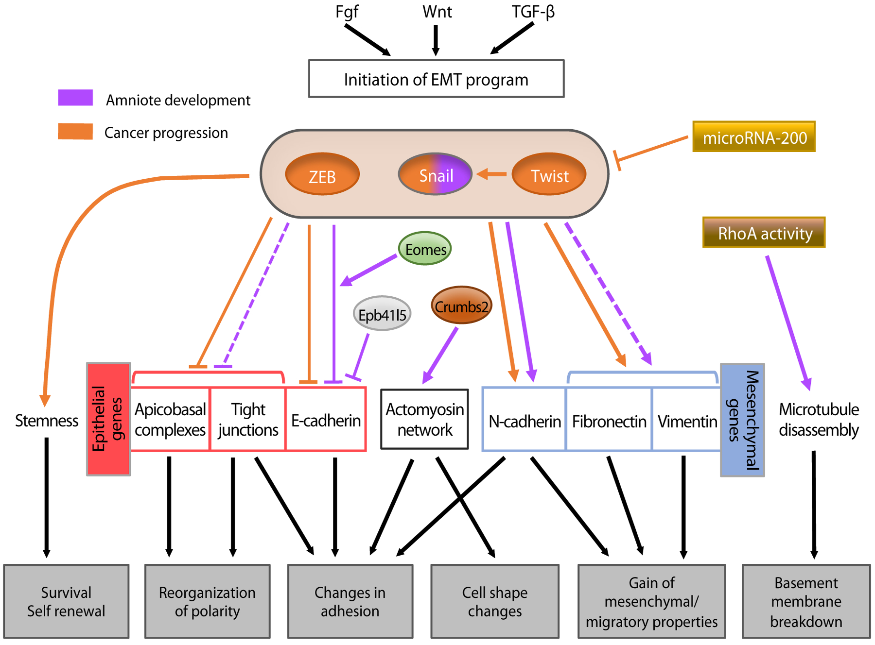
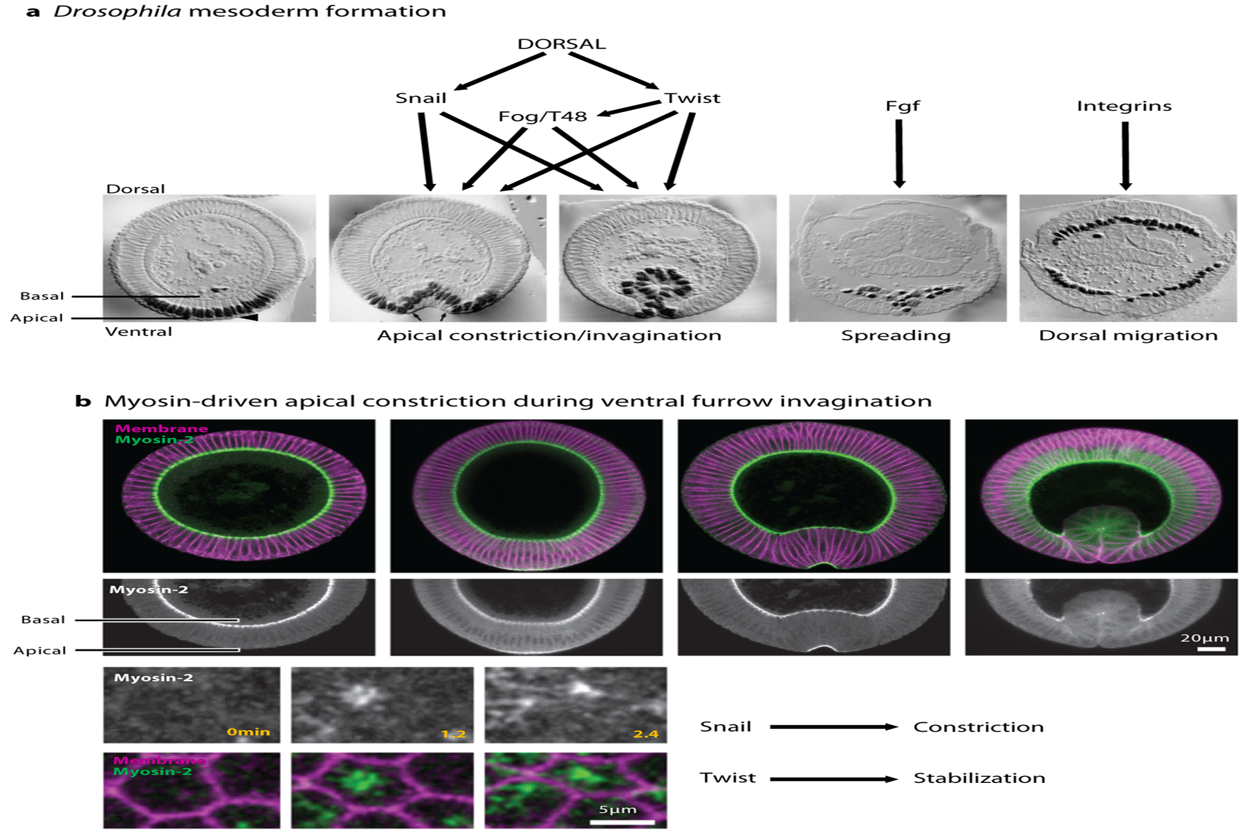
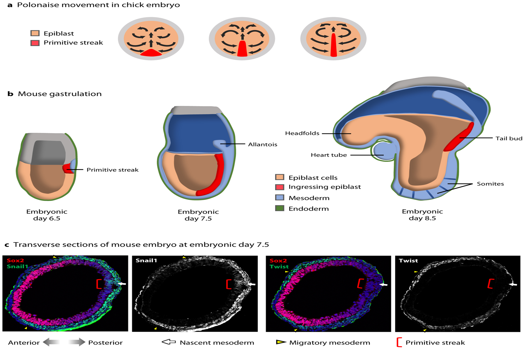
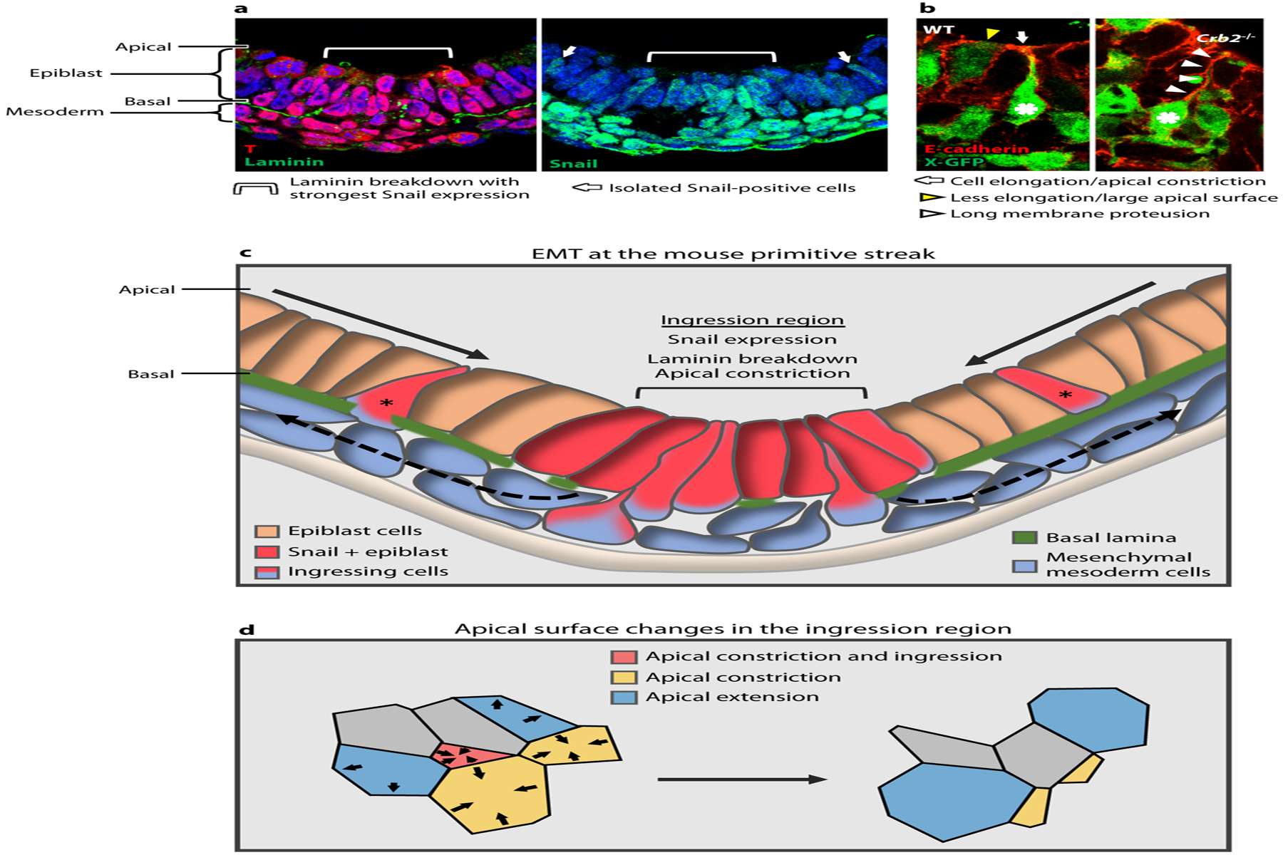
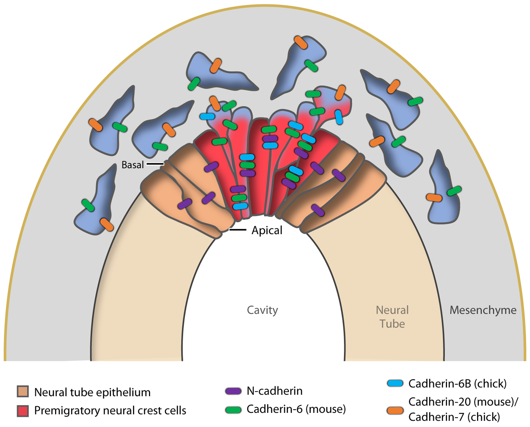
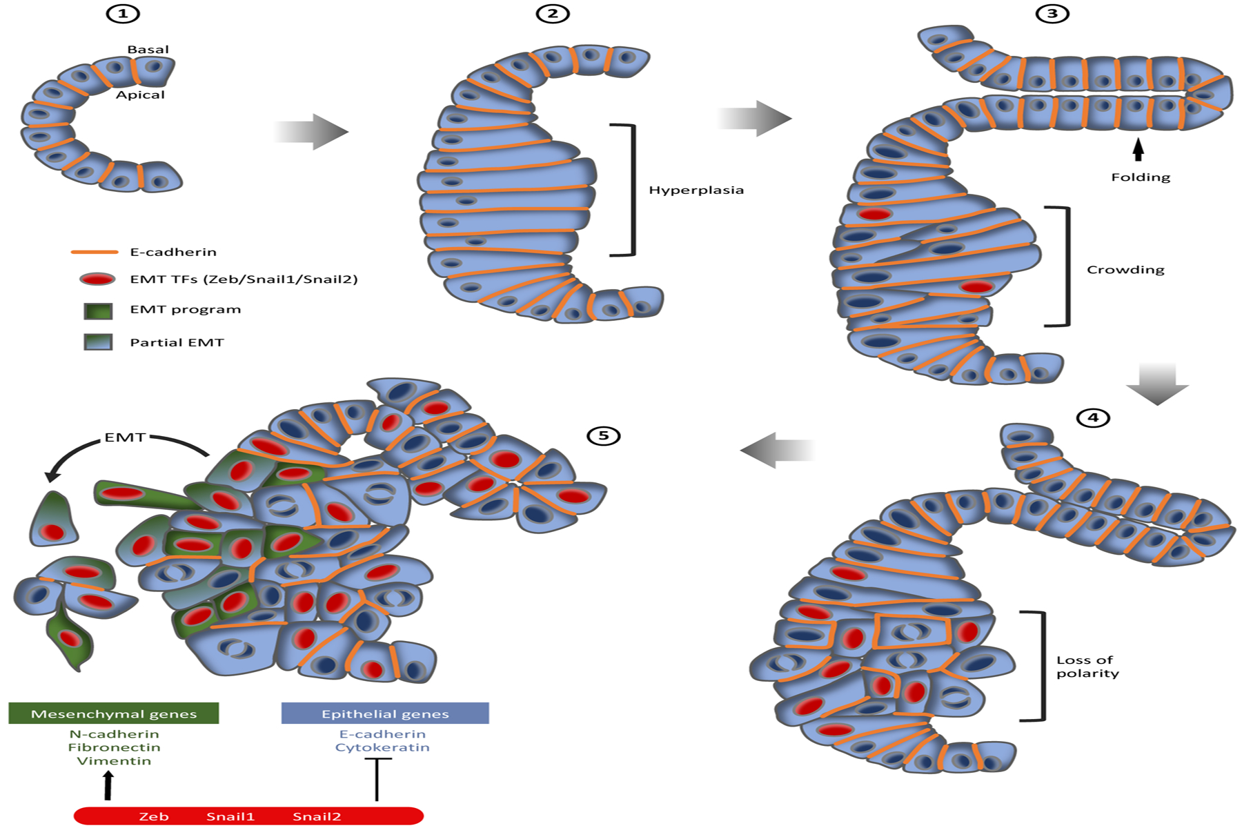
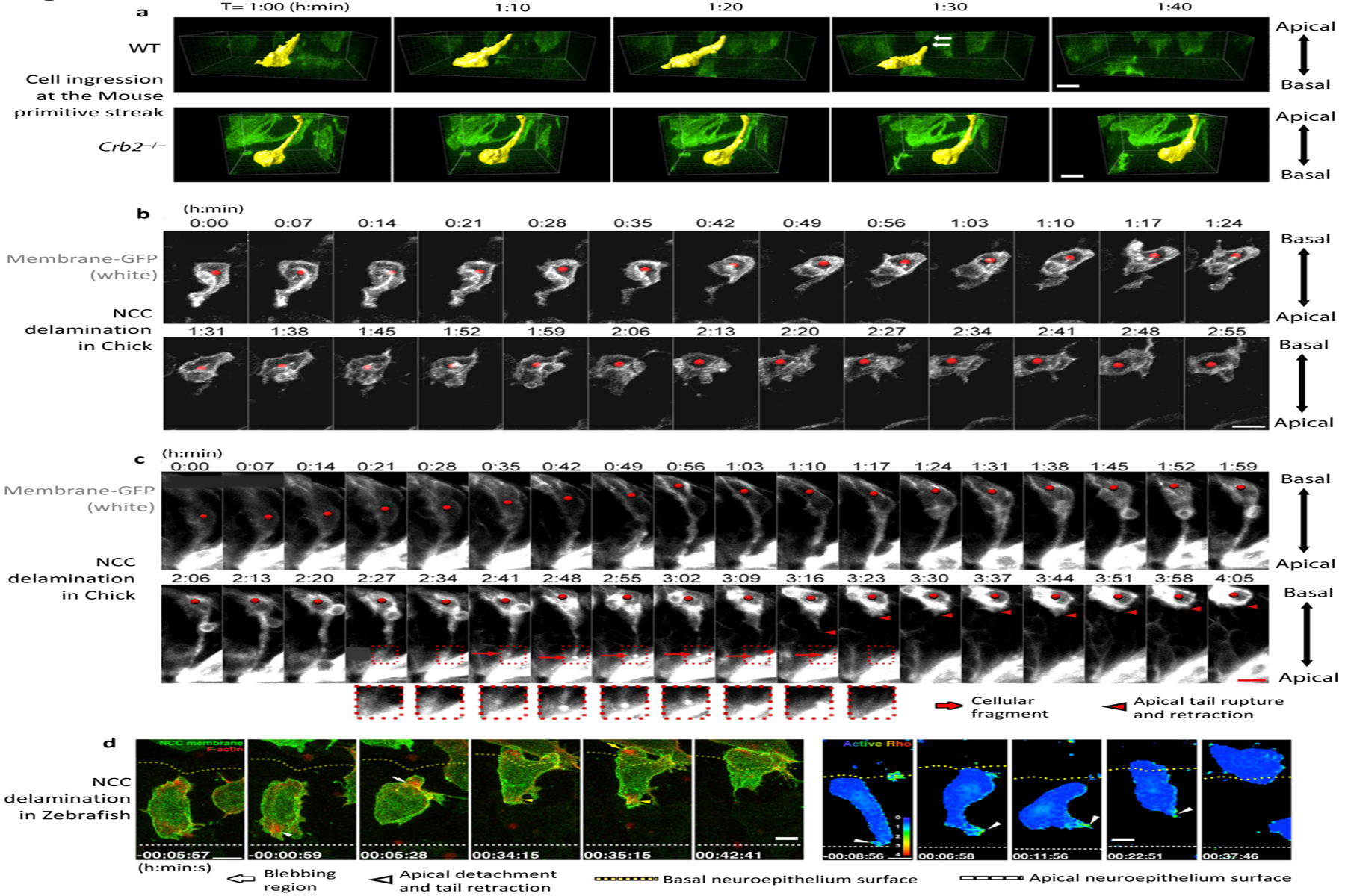
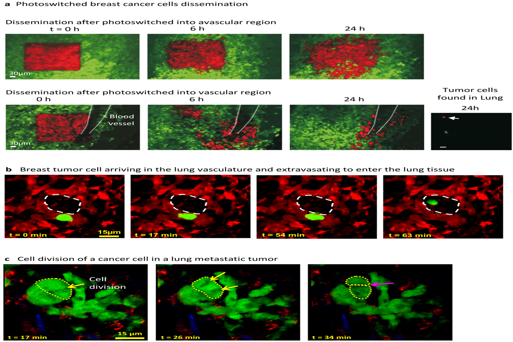
Epithelial-to-mesenchymal transitions (EMTs) are complex cellular processes where cells undergo dramatic changes in signaling, transcriptional programming, and cell shape, while directing the exit of cells from the epithelium and promoting migratory properties of the resulting mesenchyme. EMTs are essential for morphogenesis during development and are also a critical step in cancer progression and metastasis formation. Here we provide an overview of the molecular regulation of the EMT process during embryo development, focusing on chick and mouse gastrulation and neural crest development. We go on to describe how EMT regulators participate in the progression of pancreatic and breast cancer in mouse models, and discuss the parallels with developmental EMTs and how these help to understand cancer EMTs. We also highlight the differences between EMTs in tumor and in development to arrive at a broader view of cancer EMT. We conclude by discussing how further advances in the field will rely on in vivo dynamic imaging of the cellular events of EMT.

Keywords: Epithelial-to-Mesenchymal Transition; breast cancer; cancer progression; gastrulation; intravital imaging; pancreatic ductal adenocarcinoma.


Figures

Figure 1:
Figure 1:
Regulatory networks controlling epithelial-to-mesenchymal transition (EMT) during amniote development and cancer progression. Upstream signaling initiates the EMT program. Three major transcription factor families regulate the process: Snail (zinc finger proteins), Twist (a basic helix-loop-helix protein), and Zeb (zinc finger proteins that, like Twist, bind to E-box motifs). Each of these transcription factors has been implicated in cancer progression, but only Snail has been shown to play a role in developmental EMT in the mouse. The transcription factors act on downstream effectors, mainly by repressing epithelial genes and activating mesenchymal genes. Snail and Zeb mainly act as repressors and Twist acts as an activator.
Figure 2:
Figure 2:
Drosophila mesoderm formation during gastrulation. (a) Drosophila embryo sections at different stages during gastrulation, showing apical constriction and invagination of the ventral cells that will make the mesoderm, as well as spreading and dorsal migration of mesodermal cells after EMT. Black nuclei represent Twist immunostaining. Panel adapted with permission from Leptin & Grunewald (1990). (b) Myosin-2 accumulates on the apical side of ventral furrow cells and triggers apical constriction. Apico-medial accumulation of Myosin regulates pulsed apical constriction that leads to invagination. Snail controls the constriction phase and Twist stabilizes the apical surface. Figure adapted with permission from Vasquez et al. (2014).
Figure 3:
Figure 3:
Cell movements and gene expression during chick and mouse gastrulation. (a) Polonaise movement of epiblast cells associated with primitive streak elongation in the developing chick embryo, viewed from above the dorsal side. (b) Mouse gastrulation at E6.5–E8.5 from primitive streak formation, elongation, and tail bud stage, viewed from a lateral side (c) Transverse sections of E7.5 mouse embryos. Snail1 is expressed in a small population of cells in the epiblast at the primitive streak (red bracket), in the nascent mesoderm (white arrow), and in the migratory mesoderm (yellow arrowheads). Twist is not expressed at the primitive streak or in the nascent mesoderm but is turned on later in the migratory mesoderm. Images courtesy of Nitya Ramkumar.
Figure 4:
Figure 4:
Mouse primitive streak and gastrulation EMT (epithelial-to-mesenchymal transition). (a) Cross-section of the posterior side of a mouse embryo showing T staining at the primitive streak in the epiblast and mesoderm; apical epiblast is up and basal is down. Laminin breakdown occurs beneath a group 6–8 cells wide, corresponding to the strongest Snail expression in the epiblast (light blue brackets). This also corresponds to the region where 80–90% of the ingression events occur, as determined by analyzing whole-embryo time-lapse imaging (data not shown). Isolated Snail positive cells are also observed (white arrows). (b) E-cadherin staining and X-GFP at the primitive streak, show a WT embryo with an ingressing cell (asterisk) elongating and constricting its apical surface (white arrow) and neighboring cell in a different step of the process, which is less elongated and with a larger apical surface (yellow arrowhead). Crb2−/− cell moves further basally (asterisk) and retains a long membrane protrusion attached apically (white arrowheads). Panel adapted with permission from Ramkumar et al. (2016). (c) Schematic of the EMT at the primitive streak during mouse gastrulation. Epiblast cells converge toward the ingression region, which is characterized by expression of Snail, breakdown of the basal lamina, and apical constriction of individual cells. Ingression of a subset of cells at that position appears to be stochastic. Some isolated ingressions are observed outside of this region (asterisks), which may correspond to isolated Snail-positive cells. (d) In the ingression region, cells constrict their apical surfaces in an asynchronous and apparently stochastic manner.
Figure 5:
Figure 5:
EMT (epithelial-to-mesenchymal transition) and cadherin switching in neural crest cells. Premigratory neural crest cells (red) in the dorsal region of the neural tube epithelium (brown) undergo EMT, delaminate, and then migrate ventrally, switching cadherin expression.
Figure 6:
Figure 6:
The steps of pancreatic cancer progression from a normal pancreatic duct through the stages of pancreatic intraepithelial neoplasia (PanIN) and pancreatic ductal adenocarcinoma (PDAC). (❶) A normal pancreatic duct is a simple cuboidal epithelium. (❷) PanIN-1 lesions are associated with cell and tissue hyperplasia (bracket). (❸) PanIN-2 shows some nuclear enlargement, crowding (bracket), and folding of the epithelium (arrow). (❹) PanIN-3 is characterized by nuclear enlargement, loss of polarity (bracket), and the presence of frequent mitotic figures. (❺) PanIN-3 can progress into PDAC, showing loss of polarity and organization of the epithelium and partial loss of E-cadherin. Epithelial-to-mesenchymal (EMT) transcription factor (TFs) expression is observed early in PanIN-2. Cells can activate an EMT-like program or a partial-EMT program and invade surrounding tissue as isolated cells or as a cluster.
Figure 7:
Figure 7:
Dynamic imaging of ingression of cells at the primitive streak and neural crest. (a) Three-dimensional surface rendering of individual cells ingressing at the mouse primitive streak. Wild-type (WT) cells elongate in the basal direction and detach from the apical surface, whereas Crb2−/− cells move basally but remain attached to the apical surface of the epiblast. (b) Dynamics of delamination of Neural Crest Cells (NCC)in the chick embryo, where most cells detach from the apical surface, retract their apical tail, and move out of the epithelium. (c) In some cases, cells detach and leave a fragment of membrane on the apical surface. (d) Dynamics of NCC delamination in zebra fish showing F-actin accumulation in the blebbing region and at the apical tail during apical detachment and retraction, as well as Rho activity in the apical tail during detachment and retraction. In d, apical detachment is at 0 minutes. All scale bars represent 10 μm. Panels adapted with permission from (a) Ramkumar et al. (2016), (b,c) Ahlstrom & Erickson (2009), and (d) Clay & Halloran (2013.
Figure 8:
Figure 8:
Intravital imaging of breast tumor and lung metastasis. (a) Intravital visualization of an orthotopic breast tumor through a permanent mammary imaging window. Breast cancer cells in the mammary fat pad (green), when photoswitched (red) in an avascular region showed limited invasion (top), whereas when photoswitched into a vascular region, the cells infiltrated a larger area, migrated along the blood vessel, and probably intravasated (bottom); some cells were found in the lung 24 h after photoswitching (right). (b) Spontaneous metastatic cell visualized through a permanent thoracic window. A cell from an orthotopic mammary tumor (green) arriving in the lung vasculature (red), extravasating, and moving into the alveolar space (white dashed region). (c) A cluster of metastatic tumor cells in the lung showing cell division with chromosomal alignment and separation, and cytokinesis (white arrows). Panels adapted with permission from (a) Kedrin et al. (2008) and (b,c) Entenberg et al. (2018).

References

    1. Acloque H, Ocana OH, Abad D, Stern CD, Nieto MA. 2017. Snail2 and Zeb2 repress P-cadherin to define embryonic territories in the chick embryo. Development 144:649–56 - PubMed
    1. Acloque H, Ocana OH, Matheu A, Rizzoti K, Wise C, et al. 2011. Reciprocal repression between Sox3 and snail transcription factors defines embryonic territories at gastrulation. Dev. Cell 21:546–58 - PMC - PubMed
    1. Ahlstrom JD, Erickson CA. 2009. The neural crest epithelial-mesenchymal transition in 4D: a ‘tail’ of multiple non-obligatory cellular mechanisms. Development 136:1801–12 - PMC - PubMed
    1. Aiello NM, Brabletz T, Kang Y, Nieto MA, Weinberg RA, Stanger BZ. 2017. Upholding a role for EMT in pancreatic cancer metastasis. Nature 547:E7–8 - PMC - PubMed
    1. Aiello NM, Maddipati R, Norgard RJ, Balli D, Li J, et al. 2018. EMT subtype influences epithelial plasticity and mode of cell migration. Dev. Cell 45:681–95.e4 - PMC - PubMed

LinkOut - more resources