. 2021 Apr 26;24(5):102478.
doi: 10.1016/j.isci.2021.102478. eCollection 2021 May 21.

Diverse meibum lipids produced by Awat1 and Awat2 are important for stabilizing tear film and protecting the ocular surface

Affiliations

Diverse meibum lipids produced by Awat1 and Awat2 are important for stabilizing tear film and protecting the ocular surface

Megumi Sawai et al. iScience. .

Erratum in

Abstract

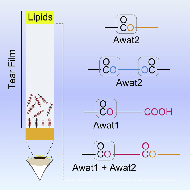
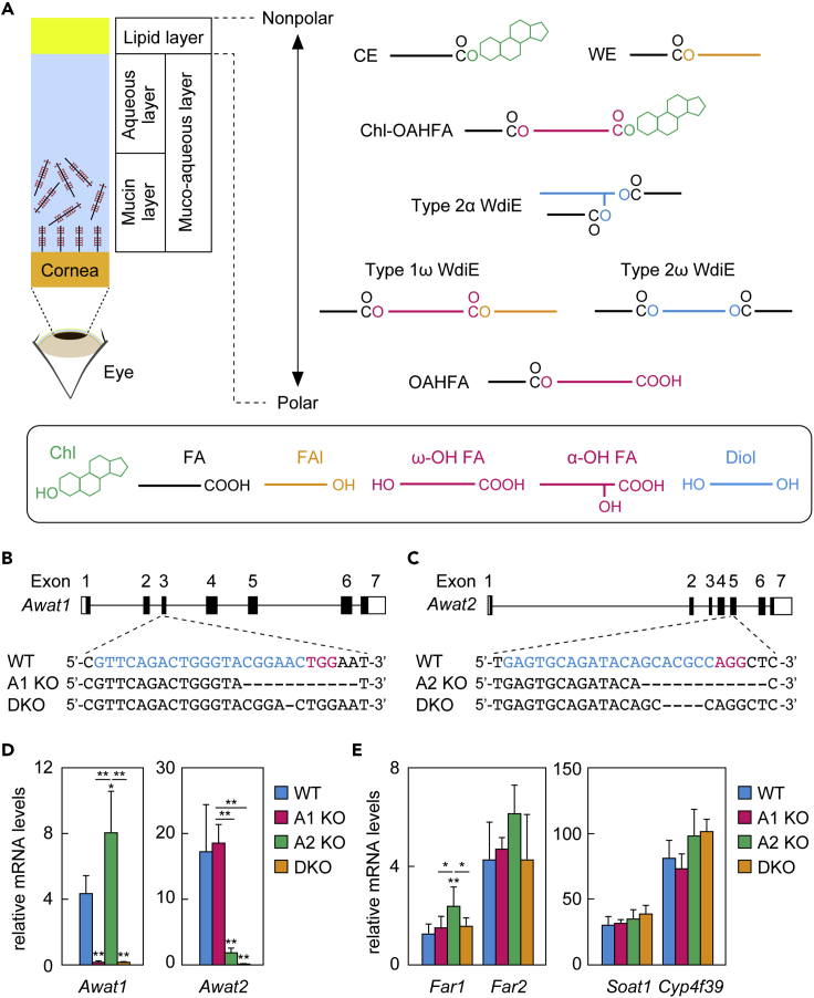
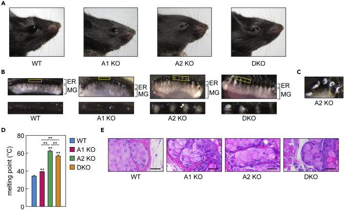
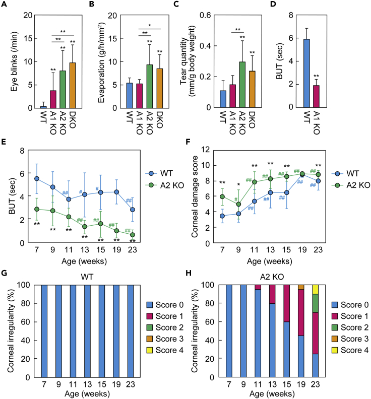
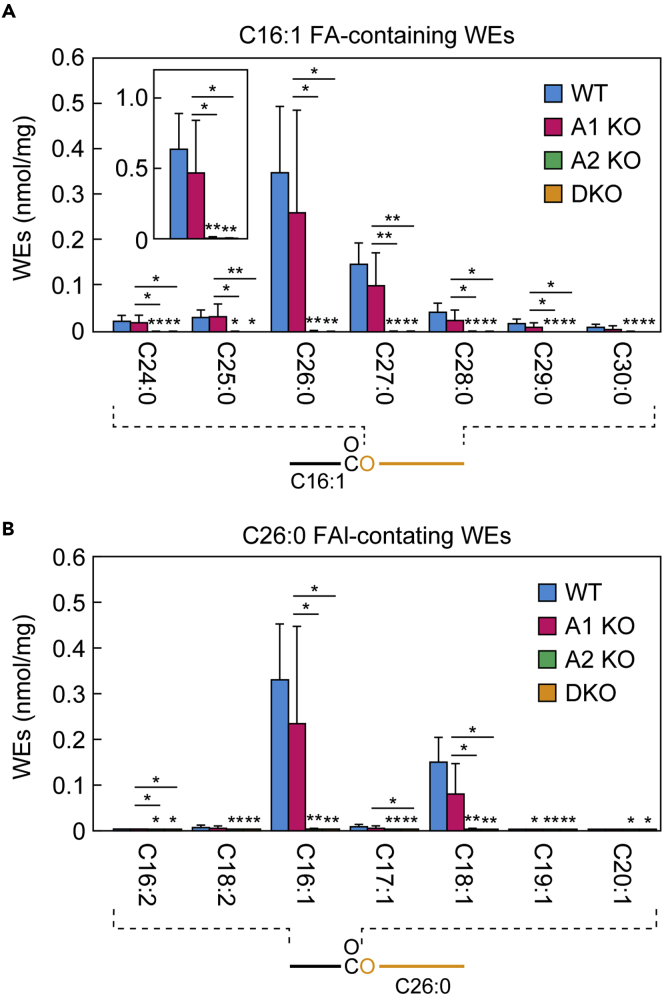
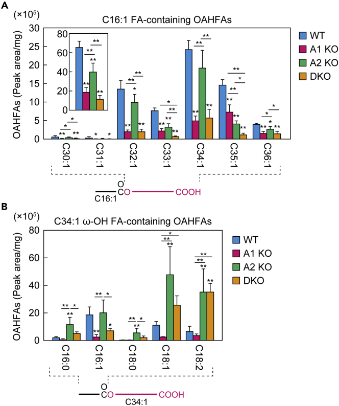
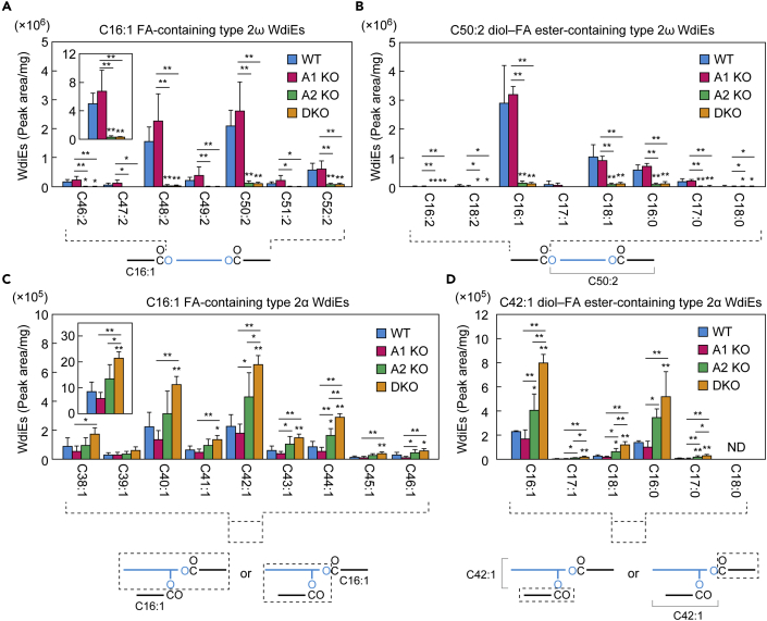
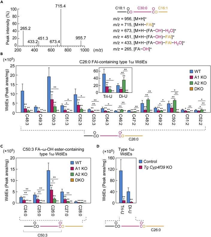
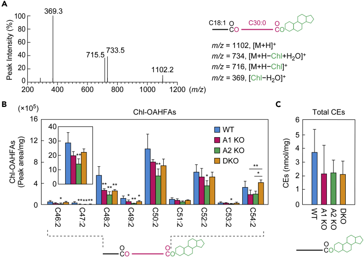
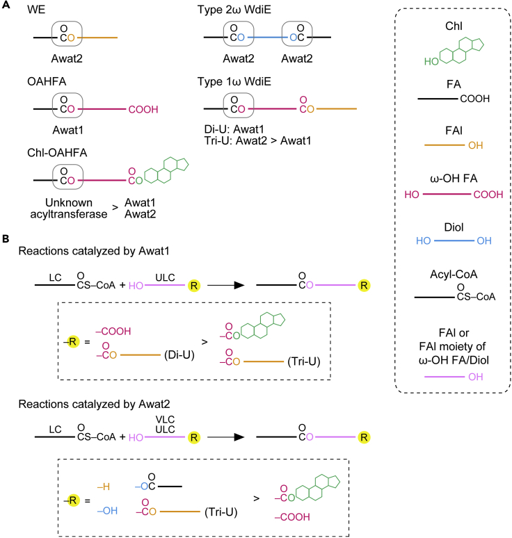
A lipid layer consisting of meibum lipids exists in the tear film and functions in preventing dry eye disease. Although the meibum lipids include diverse lipid classes, the synthesis pathway and role of each class remain largely unknown. Here, we created single and double knockout (KO and DKO, respectively) mice for the two acyl-CoA wax alcohol acyltransferases (Awat1 and Awat2) and investigated their dry eye phenotypes and meibum lipid composition. Awat2 KO and DKO mice exhibited severe dry eye with meibomian gland dysfunction, whereas Awat1 KO mice had mild dry eye. In these mice, specific meibum lipid classes were reduced: (O-acyl)-ω-hydroxy fatty acids and type 1ω wax diesters in Awat1 KO mice, wax monoesters and types 1ω and 2ω wax diesters in Awat2 KO mice, and most of these in DKO mice. Our findings reveal that Awat1 and Awat2 show characteristic substrate specificity and together produce diverse meibum lipids.


Conflict of interest statement

The authors declare no competing interests.

Figures

None
Graphical abstract
Figure 1
Figure 1
Generation of Awat1 KO, Awat2 KO, and Awat1 Awat2 DKO mice (A) Schematic illustration of the eye, cornea, and tear film and the simplified structures of the major meibum lipids. (B and C) The gene structures (black, coding regions; white, untranslated regions) of Awat1 (B) and Awat2 (C) and the nucleotide sequences around the guide RNA target sequences. The blue and red nucleotides in the WT sequence represent the target sequence and the protospacer-adjacent motif sequence, respectively. (D and E) Total RNAs were prepared from the meibomian glands of 6-week-old WT (n = 4), Awat1 KO (n = 4), Awat2 KO (n = 4), and DKO (n = 4) mice and subjected to real-time quantitative RT-PCR using specific primers for Awat1 (D), Awat2 (D), Far1 (E), Far2 (E), Soat1 (E), Cyp4f39 (E), or the housekeeping gene Hprt (D and E). Values presented are mean (±SD) quantities of each mRNA relative to those of Hprt. Significant differences from the WT mice (asterisks above columns) and among the mutants (asterisks above horizontal lines) are indicated (∗p < 0.05; ∗∗p < 0.01; Tukey's test). A1 KO, Awat1 KO; A2 KO, Awat2 KO.
Figure 2
Figure 2
Awat1 and/or Awat2 deficiency causes plugging of the meibomian gland orifices (A) Photographs of 6-week-old WT, Awat1 KO, Awat2 KO, and Awat1 Awat2 DKO mice. (B) Upper eyelids from 6-week-old WT, Awat1 KO, Awat2 KO, and DKO mice, photographed under a light microscope. The lower images are magnified views of the yellow rectangles in the upper images and show the meibomian gland (MG) orifices. ER, eyelid rim. (C) The upper eyelid of a 6-week-old Awat2 KO mouse after the meibomian glands were subjected to pressure, causing them to extrude meibum from the orifices. (D) The melting point of meibum lipids extruded from the meibomian gland orifices was measured in 6-week-old WT (n = 3), Awat1 KO (n = 3), Awat2 KO (n = 3), and DKO (n = 3) mice. Values presented are means ± SD. Significant differences from WT mice (asterisks above columns) and among the mutants (asterisks above horizontal lines) are indicated (∗∗p < 0.01; Tukey's test). (E) Paraffin sections of the meibomian glands in 6-week-old WT, Awat1 KO, Awat2 KO, and DKO mice were stained with hematoxylin and eosin. The bright-field images were photographed under a light microscope. Scale bar, 25 μm. A1 KO, Awat1 KO; A2 KO, Awat2 KO.
Figure 3
Figure 3
Awat1 or Awat2 deficiency causes dry eye phenotypes (A–C) Blink frequency (A), water evaporation from the ocular surface (B), and tear quantity (C) were measured in 6-week-old WT, Awat1 KO, Awat2 KO, and Awat1 Awat2 DKO mice. Values presented are means ± SD. The number of mice of each line examined was as follows: WT, n = 30 (A) or n = 28 (B and C); Awat1 KO, n = 14 (A) or n = 11 (B and C); Awat2 KO, n = 14 (A and C) or n = 13 (B); DKO, n = 17 (A) or n = 16 (B and C). Significant differences from the WT mice (asterisks above columns) and among the mutants (asterisks above horizontal lines) are indicated (∗∗p < 0.01; Tukey-Kramer test). (D) BUT was measured in 6-week-old WT (n = 3) and Awat1 KO (n = 3) mice. Values presented are means ± SD. Significant differences from the WT mice are indicated (∗∗p < 0.01; Student's t-test). (E–H) BUT (E), corneal damage score (F), and corneal surface irregularity score (G and H) were measured in WT (n = 10) (E–G) and Awat2 KO (n = 10) (E, F, and H) mice. Experiments were performed from the age of 7 weeks to 23 weeks, and measurements were performed on both eyes. (E and F) Values presented are means ± SD, and significant differences from WT mice of the same age (∗p < 0.05; ∗∗p < 0.01; Student's t-test) and from mice of the same genotype at 7 weeks old (#p < 0.05; ##p < 0.01; Dunnett's test) are indicated. (G and H) The proportion of the mice that had each score (0–4) at each age. A1 KO, Awat1 KO; A2 KO, Awat2 KO.
Figure 4
Figure 4
Awat2 is involved in WE production Lipids were extracted from the meibomian glands of 6-week-old WT (n = 5), Awat1 KO (n = 5), Awat2 KO (n = 5), and Awat1 Awat2 DKO (n = 5) mice, and WEs were analyzed using LC-MS/MS. (A) Quantities of the major WEs (nmol/mg tissue) composed of a C16:1 FA moiety and a saturated FAl moiety with a chain length of C24–C30. Inset shows the total quantity of WEs composed of a C16:1 FA moiety and a saturated FAl moiety with a chain length of C16–C36. (B) Quantities of WEs (nmol/mg tissue) containing one of the indicated FA moieties and a C26:0 FAl moiety. Values presented are means ± SD, and significant differences from the WT mice (asterisks above columns) and among the mutants (asterisks above horizontal lines) are indicated (∗p < 0.05; ∗∗p < 0.01; Tukey's test). The simplified structure of a WE with the analyzed moiety (FAl or FA) indicated is shown below each graph. Data for all WEs measured are provided in Table S1. A1 KO, Awat1 KO; A2 KO, Awat2 KO.
Figure 5
Figure 5
Awat1 is involved in OAHFA production Lipids were extracted from the meibomian glands of 6-week-old WT (n = 4), Awat1 KO (n = 4), Awat2 KO (n = 4), and Awat1 Awat2 DKO (n = 4) mice. After derivatization with N-(4-aminomethylphenyl)pyridinium, OAHFAs were analyzed using LC-MS/MS. (A) Quantities of OAHFAs (peak area/mg tissue) composed of a C16:1 FA moiety and a monounsaturated ω-OH FA moiety with a chain length of C30–C36. Inset shows the total quantities of OAHFAs composed of a C16:1 FA moiety and a monounsaturated ω-OH FA moiety with a chain length of C16–C36. (B) Quantities of OAHFAs (peak area/mg tissue) containing one of the indicated FA moieties and a C34:1 ω-OH FA moiety. Values presented are means ± SD, and significant differences from the WT mice (asterisks above columns) and among the mutants (asterisks above horizontal lines) are indicated (∗p < 0.05; ∗∗p < 0.01; Tukey's test). The simplified structure of an OAHFA with the analyzed moiety (ω-OH FA or FA) indicated is shown below each graph. Data for all OAHFAs measured are provided in Table S2. A1 KO, Awat1 KO; A2 KO, Awat2 KO.
Figure 6
Figure 6
Awat2 is involved in type 2ω WdiE production Lipids were extracted from the meibomian glands of 6-week-old WT (n = 4), Awat1 KO (n = 4), Awat2 KO (n = 4), and Awat1 Awat2 DKO (n = 4) mice, and type 2ω WdiEs (A and B) and type 2α WdiEs (C and D) were analyzed using LC-MS/MS. (A) Quantities of type 2ω WdiEs (peak area/mg tissue) composed of a C16:1 FA moiety and a di-unsaturated diol-FA ester moiety with a chain length of C46–C52. Inset shows the total quantities of type 2ω WdiEs composed of a C16:1 FA moiety and a di-unsaturated diol-FA ester moiety with a chain length of C32–C54. (B) Quantities of type 2ω WdiEs (peak area/mg tissue) composed of one of the indicated FA moieties and a C50:2 diol-FA ester moiety. (C) Quantities of type 2α WdiEs (peak area/mg tissue) composed of a C16:1 FA moiety and a monounsaturated diol-FA ester moiety with a chain length of C38–C46. Inset shows the total quantities of type 2α WdiEs composed of a C16:1 FA moiety and a monounsaturated diol-FA ester moiety with a chain length of C32–C54. (D) Quantities of type 2α WdiEs (peak area/mg tissue) composed of one of the indicated FA moieties and a C42:1 diol-FA ester moiety. Values presented are means ± SD, and significant differences from the WT mice (asterisks above columns) and among the mutants (asterisks above horizontal lines) are indicated (∗p < 0.05; ∗∗p < 0.01; Tukey's test). The simplified structure of a WdiE with the analyzed moiety (diol-FA ester or FA) indicated is shown below each graph. Data for all type 2ω and 2α WdiEs measured are provided in Tables S3 and S4, respectively. A1 KO, Awat1 KO; A2 KO, Awat2 KO; ND, not detected.
Figure 7
Figure 7
Awat1 and Awat2 are differentially involved in di-unsaturated and tri-unsaturated type 1ω WdiE production (A) Product ion scanning of the chemically synthesized type 1ω WdiE standard (C18:1 FA + ω-OH C30:0 FA + C16:1 FAl) was performed using LC-MS/MS, by selecting the [M + H]+ ion with m/z = 955.9 as a precursor. The MS spectrogram and the predicted product ions are shown. The synthesis scheme for the type 1ω WdiE standard is provided in Figure S3. (B–D) Lipids were extracted from the meibomian glands of 6-week-old WT (n = 4), Awat1 KO (n = 4), Awat2 KO (n = 4), and Awat1 Awat2 DKO (n = 4) mice (B and C), or from the meibomian glands of 12-month-old Tg Cyp4f39 KO (n = 3) and their control (n = 3) mice (D), and type 1ω WdiEs were analyzed using LC-MS/MS. (B) Quantities of type 1ω WdiEs (peak area/mg tissue) composed of a C26:0 ULCFAl moiety and one of the indicated di-unsaturated or tri-unsaturated FA-ω-OH FA ester moieties. Inset shows the total quantities of type 1ω WdiEs composed of a C26:0 ULCFAl moiety and a di-unsaturated or tri-unsaturated FA-ω-OH FA ester moiety with a chain length of C32–C54. (C) Quantities of type 1ω WdiEs (peak area/mg tissue) composed of a saturated FAl moiety with a chain length of C24–C28 and a C50:3 FA-ω-OH FA ester moiety. (D) The total amounts of type 1ω WdiEs (peak area/mg tissue) composed of a C26:0 ULCFAl moiety and a di-unsaturated or tri-unsaturated FA-ω-OH FA ester moiety of C32–C54 chain lengths. Values presented are means ± SD, and significant differences from the WT (B and C) or control mice (D) (asterisks above columns) and among the mutants (asterisks above horizontal lines) are indicated (∗p < 0.05; ∗∗p < 0.01; Tukey's test [B and C] or Student's t-test [D]). The simplified structure of a type 1ω WdiE with the analyzed moiety (FA-ω-OH FA ester or FAl) indicated is shown below each graph. Data for all type 1ω WdiEs measured are provided in Tables S5 and S6. A1 KO, Awat1 KO; A2 KO, Awat2 KO. Di-U, di-unsaturated; Tri-U, tri-unsaturated.
Figure 8
Figure 8
Awat1 and Awat2 are partially involved in Chl-OAHFA production (A) Product ion scanning of the chemically synthesized Chl-OAHFA standard (C18:1 FA + ω-OH C30:0 FA + Chl) was performed using LC-MS/MS by selecting the [M + H]+ ion with m/z = 1102.1 as a precursor. The MS spectrogram and the predicted product ions are shown. The synthesis scheme for the Chl-OAHFA standard is provided in Figure S4. (B) Lipids were extracted from the meibomian glands of 6-week-old WT (n = 4), Awat1 KO (n = 4), Awat2 KO (n = 4), and Awat1 Awat2 DKO (n = 4) mice, and Chl-OAHFAs were analyzed using LC-MS/MS. Quantities of Chl-OAHFAs (peak area/mg tissue) containing a di-unsaturated OAHFA moiety with a chain length of C46–C54 are shown. Inset shows the total quantities of Chl-OAHFAs containing a di-unsaturated OAHFA moiety with a chain length of C32–C54. The simplified structure of a Chl-OAHFA with the analyzed moiety (OAHFA) indicated is shown below the graph. (C) Lipids were extracted from the meibomian glands of 6-week-old WT (n = 5), Awat1 KO (n = 5), Awat2 KO (n = 5), and DKO (n = 5) mice, and CEs were analyzed using LC-MS/MS. Total quantities of CEs (nmol/mg tissue) containing a saturated or monounsaturated FA moiety with a chain length of C16–C36 are shown. Values presented are means ± SD, and significant differences from the WT mice (asterisks above columns) and among the mutants (asterisks above horizontal lines) are indicated (∗p < 0.05; ∗∗p < 0.01; Tukey's test). Data for all Chl-OAHFAs and CEs measured are provided in Tables S7 and S8, respectively. A1 KO, Awat1 KO; A2 KO, Awat2 KO.
Figure 9
Figure 9
Model for the substrate specificity of Awat1 and Awat2 in the production of diverse meibum lipids (A) Models for the involvement of Awat1 and Awat2 in the formation of ester bonds (in gray boxes) to produce the indicated meibum lipids. Inequality signs denote the relative contribution of the enzymes involved. (B) Substrate specificity of Awat1 and Awat2 toward FAl and FAl derivatives. Di-U, di-unsaturated; Tri-U, tri-unsaturated.

Similar articles

Cited by

References

    1. Bron A.J., de Paiva C.S., Chauhan S.K., Bonini S., Gabison E.E., Jain S., Knop E., Markoulli M., Ogawa Y., Perez V. TFOS DEWS II pathophysiology report. Ocul. Surf. 2017;15:438–510. - PubMed
    1. Bron A.J., Tiffany J.M., Gouveia S.M., Yokoi N., Voon L.W. Functional aspects of the tear film lipid layer. Exp. Eye Res. 2004;78:347–360. - PubMed
    1. Butovich I.A. The Meibomian puzzle: combining pieces together. Prog. Retin. Eye Res. 2009;28:483–498. - PMC - PubMed
    1. Butovich I.A. Meibomian glands, meibum, and meibogenesis. Exp. Eye Res. 2017;163:2–16. - PMC - PubMed
    1. Butovich I.A., Wilkerson A., Bhat N., McMahon A., Yuksel S. On the pivotal role of Elovl3/ELOVL3 in meibogenesis and ocular physiology of mice. FASEB J. 2019;33:10034–10048. - PMC - PubMed