. 2021 May 1;24(5):102493.
doi: 10.1016/j.isci.2021.102493. eCollection 2021 May 21.

Replication protein A plays multifaceted roles complementary to specialized helicases in processing G-quadruplex DNA

Affiliations

Replication protein A plays multifaceted roles complementary to specialized helicases in processing G-quadruplex DNA

Yi-Ran Wang et al. iScience. .

Abstract

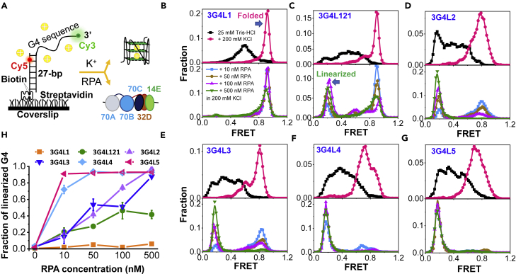
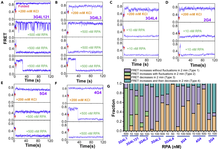
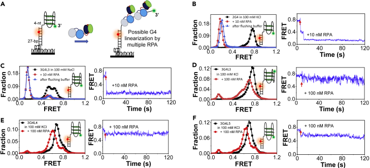
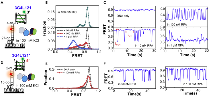
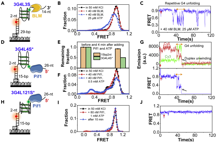
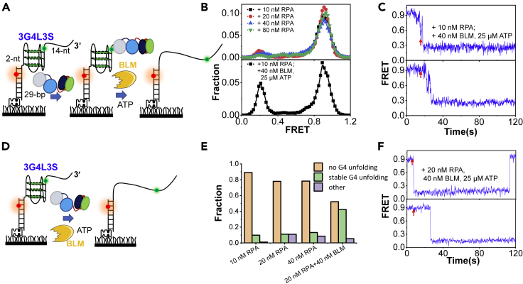
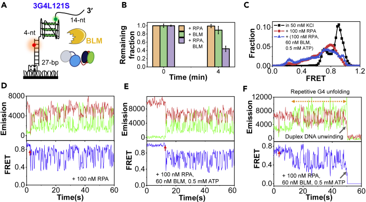
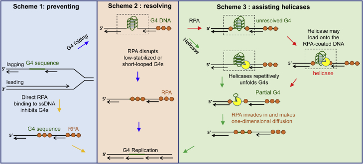
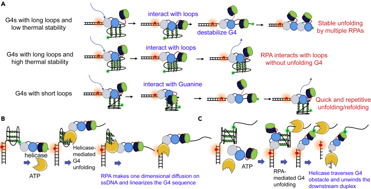
G-quadruplexes (G4s) are non-canonical DNA structures with critical roles in DNA metabolisms. To resolve those structures that can cause replication fork stalling and genomic instability, single-stranded DNA-binding proteins and helicases are required. Here, we characterized the interplay between RPA and helicases on G4s using single-molecule FRET. We first discovered that human RPA efficiently prevents G4 formation by preempting ssDNA before its folding. RPA also differentially interacts with the folded G4s. However, helicases such as human BLM and yeast Pif1 have different G4 preferences from RPA mainly based on loop lengths. More importantly, both RPA and these helicases are required for the stable G4 unfolding, as RPA promotes helicase-mediated repetitive unfolding into durative linear state. Furthermore, BLM can traverse G4 obstacles temporarily disrupted by RPA and continue to unwind downstream duplex. We finally proposed the mechanisms underlying above functions of RPA in preventing, resolving, and assisting helicases to eliminate G4s.

Keywords: Molecular biology; Molecular structure.


Conflict of interest statement

The authors declare no competing interests.

Figures

None
Graphical abstract
Figure 1
Figure 1
RPA significantly suppresses the folding of G-rich DNA sequences into G4 structures (A) Schematic diagram of the Cy3- and Cy5-labeled DNA harboring G4 motif. K+ will drive G4 sequence folding into G4 structure; however, on the opposite, the tight occupation by RPA (a trimer composed of 70A, 70B, and 70C domains in the 70 kD subunit; the 32 kD subunit; and the 14 kD subunit) may maintain G4 motif in the linearized state. (B–G) The upper panels are the FRET distributions of G4 motifs in 25 mM Tris-HCl before and 4 min after the addition of 200 mM KCl with 5 mM MgCl2. The lower panels are the FRET distributions after the addition of RPA in 200 mM KCl and 5 mM MgCl2 to the G4 DNA substrates originally prepared in 25 mM Tris-HCl. (H) The fractions of linearized G4s were obtained from the FRET peaks at ∼E0.2. The error bars were obtained from at least three repetitive experiments. Data are presented as mean ± SEM.
Figure 2
Figure 2
RPA prevents the formation of G4 structures by preempting ssDNA before its folding (A–F) The representative FRET traces of G4 DNA substrates prepared in 25 mM Tris-HCl, pH 7.5 upon the addition of 200 mM KCl and 5 mM MgCl2 (panel 1) and RPA in 200 mM KCl with 5 mM MgCl2 (panels 2–4). The red arrow indicates the addition of ions or proteins, i.e., the start of the flow to the ∼25 μL flow cell. (G) The fractions of FRET traces showing different types of changes upon the addition of RPA. All the single-molecule traces in each experiment were examined and classified into different types.
Figure 3
Figure 3
The interaction between RPA and different preformed G4 structures (A) The schematic diagram of the experimental design. Preformed G4 substrates were anchored at the coverslip surface, and then different concentrations of RPA were added to unfold the G4 structures. For G4 substrates prepared in 100 mM KCl (or 100 mM NaCl), the reaction buffer of RPA is composed of 100 mM KCl (or 100 mM NaCl) and 5 mM MgCl2. (B and C) The left panels are the FRET distributions of 2G4 in 100 mM KCl and 3G4L3 in 100 mM NaCl before and 4 min after the addition of 10 nM RPA. Washing the channels with buffer leads to little difference in the FRET distributions. The right panels are the representative traces. The red arrow indicates the addition of RPA. (D–F) The left panels are the FRET distributions of 3G4L3, 3G4L4, and 3G4L5 in 100 mM KCl before and 4 min after the addition of 100 nM RPA. The right panels are the representative traces.
Figure 4
Figure 4
RPA can rapidly and repetitively disrupt the G4 structures with very short loops (A) Schematic diagram of the experimental design. The G4 substrate has three loops of 1-, 2-, and 1-nt, respectively. (B) The FRET distributions of 3G4L121 before and 4 min after the addition of varying concentrations of RPA. (C) The representative FRET traces. tG4 denotes the duration time of G4 folding in the presence of RPA. tnG4 denotes the duration time of G4 unfolding. (D) The interaction of RPA with G4 structure being placed at the 5′-end of the duplex DNA. (E) The FRET distributions of 3G4L121∗ before and 4 min after the addition of RPA. (F) The FRET traces in 50–100 nM RPA.
Figure 5
Figure 5
The helicases-mediated unfolding of G4 DNA structures with different loop lengths (A) The 3-layered G4 substrate with three 3-nt loops for the 3′–5′ helicase. The ssDNA tail and stem duplex are 14-nt and 29-bp, respectively. (B) FRET distributions before and after the addition of 40 nM BLM and 25 μM ATP in 25 mM Tris-HCl, 50 mM KCl, and 5 mM MgCl2. Adding only 40 nM BLM without ATP has no detectable effect on the FRET distribution of G4. (C) The representative trace. (D) The 3-layered G4 substrate with three 4-nt loops for the 5′–3′ helicase. The ssDNA tail and stem duplex are 26-nt and 15-bp, respectively. (E) The remaining fractions of 3G4L4S∗ and 15bp2nt on the coverslip surface before and 4 min after the addition of 40 nM Pif1 and 0.5 mM ATP in 25 mM Tris-HCl, 50 mM KCl, and 5 mM MgCl2. Pif1 cannot unwind the duplex directly from the 2-nt ssDNA linker. The error bars were obtained from at least three repetitive experiments. Data are presented as mean ± SEM. (F) FRET distributions of the remaining 3G4L4S∗ molecules on coverslip before and after the addition of Pif1. Adding only 40 nM Pif1 without ATP has no obvious effect on the FRET distribution of G4. (G) The representative traces. (H) The 3-layered G4 substrate with three loops of 1-, 2-, and 1-nt for Pif1 helicase. (I) FRET distributions of 3G4L121S∗ before and 4 min after the addition of 80 nM Pif1 and 1 mM ATP. Even after 10 min, there is no obvious G4 unfolding. (J) The representative trace. No G4 unfolding can be observed.
Figure 6
Figure 6
RPA promotes BLM-mediated G4 unfolding into the durative linearized state (A) RPA was incubated with the substrate 3G4L3S first, and then BLM and ATP were introduced in the reaction buffer containing 50 mM KCl and 5 mM MgCl2. (B) The FRET distributions of 3G4L3S in different concentrations of RPA and after the addition of BLM to the RPA-bound 3G4L3S. (C) Representative traces after the addition of BLM and ATP to the RPA-bound 3G4L3S. (D) RPA and BLM were added simultaneously to the 3G4L3S. (E) The fractions of different types of traces at different concentrations of RPA and BLM with 25 μM ATP. (F) Representative traces of 3G4L3S in the mixture of 20 nM RPA, 40 nM BLM, and 25 μM ATP.
Figure 7
Figure 7
RPA assists BLM to overcome the G4 structure and continue to unwind the downstream duplex DNA (A) The schematic diagram of the substrate. The ssDNA tail and duplex stem are 14-nt and 27-bp, respectively. There is a 4-nt linker between G4 and duplex. (B) The remaining fractions of duplexes on coverslip before and 4 min after the addition of 100 nM RPA, 60 nM BLM, and 0.5 mM ATP and the mixture of 100 nM RPA, 60 nM BLM, and 0.5 mM ATP. The error bars were obtained from at least three repetitive experiments. Data are presented as mean ± SEM. (C) The FRET distributions of DNA molecules on coverslips before and after the addition of proteins. (D) Selected trace after the addition of 100 nM RPA. (E and F) Selected traces after the addition of 100 nM RPA, 60 nM BLM, and 0.5 mM ATP.
Figure 8
Figure 8
The proposed mechanisms of RPA in eliminating G4 structures DNA replication was taken for example. G4 structures can be formed in both the leading and lagging strands. Here the G4 in the lagging strand was shown. In scheme 1, RPA quickly and directly binds to the exposed G4 sequence, directly inhibiting its folding. Once the G4 sequence escapes RPA, it may well fold into G4 structures. In scheme 2, RPA is able to constantly disrupt the low stabilized G4s or repetitively unfold the short-looped G4s. However, RPA is poor at resolving G4s with long loops and high stability. Then in scheme 3, RPA assists the specialized helicases to remove those G4 obstacles no matter RPA coats the adjacent ssDNA at first or the helicase binds to the G4 substrate at first.
Figure 9
Figure 9
The possible interacting modes of RPA with G4 structures in the absence or presence of helicases (A) The proposed mechanisms of the interaction between RPA and different types of G4 DNA. First, for G4s with long loops (≥3-nt) and low thermal stability (Tm approximately or lower than ∼50°C), RPA may first load onto the substrate at the 4-nt linker by 70A or 70B domains. Then the trimerization core dynamically interacts with the loops, leading to the destabilization of the G4 structure. In addition, the G4 structures can be finally stably linearized by multiple RPA molecules. Second, for G4s with long loops (≥3-nt) and high thermal stability (Tm obviously higher than ∼50°C), the trimerization core predominantly interacts with the loops and the G4 structures are still intact. Third, for G4s with short loops (<3-nt), the trimerization core interacts with the Guanines in the G-tetrads, resulting in the complete or partial G4 unfolding. However, the unfolded G4s have a very strong tendency to refold back. Therefore, quick and repetitive unfolding/refolding can be observed. (B) The proposed functions of RPA in helicases-mediated G4 unfolding. Once the G4 structure was unfolded by helicases into intermediate or ssDNA states, RPA may rapidly invade into the exposed strand by one-dimensional diffusion on the ssDNA and finally, maintain the G4 strand at the linearized state. (C) Once the G4 was unfolded by RPA, helicase was able to traverse the G4 obstacle and further unwind the downstream duplex DNA.

References

    1. Audry J., Maestroni L., Delagoutte E., Gauthier T., Nakamura T.M., Gachet Y., Saintome C., Geli V., Coulon S. RPA prevents G-rich structure formation at lagging-strand telomeres to allow maintenance of chromosome ends. EMBO J. 2015;34:1942–1958. - PMC - PubMed
    1. Bochman M.L., Paeschke K., Zakian V.A. DNA secondary structures: stability and function of G-quadruplex structures. Nat. Rev. Genet. 2012;13:770–780. - PMC - PubMed
    1. Boule J.B., Zakian V.A. The yeast Pif1p DNA helicase preferentially unwinds RNA DNA substrates. Nucleic Acids Res. 2007;35:5809–5818. - PMC - PubMed
    1. Byrd A.K., Raney K.D. Structure and function of Pif1 helicase. Biochem. Soc. Trans. 2017;45:1159–1171. - PMC - PubMed
    1. Capra J.A., Paeschke K., Singh M., Zakian V.A. G-quadruplex DNA sequences are evolutionarily conserved and associated with distinct genomic features in Saccharomyces cerevisiae. PLoS Comput. Biol. 2010;6:e1000861. - PMC - PubMed

LinkOut - more resources