

doi: 10.1016/j.hroo.2020.03.001.
eCollection 2020 Apr.
His-bundle pacing is the best approach to physiological pacing
Affiliations
- PMID: 34113860
- PMCID: PMC8183870
- DOI: 10.1016/j.hroo.2020.03.001
Item in Clipboard
His-bundle pacing is the best approach to physiological pacing
Heart Rhythm O2.
.
No abstract available
Keywords: Biventricular pacing; Cardiac resynchronization therapy; His-bundle pacing; Left bundle branch block; Pacemaker; Right bundle branch block.
Figures
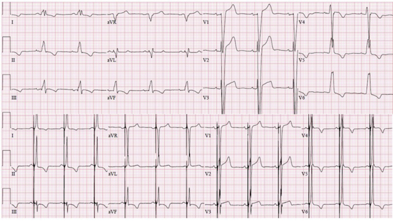
Complete correction of wide QRS (top: left bundle branch block) with His-bundle pacing (HBP), restoring physiological conduction through intrinsic activation of the His–Purkinje system (bottom: corrected HBP).
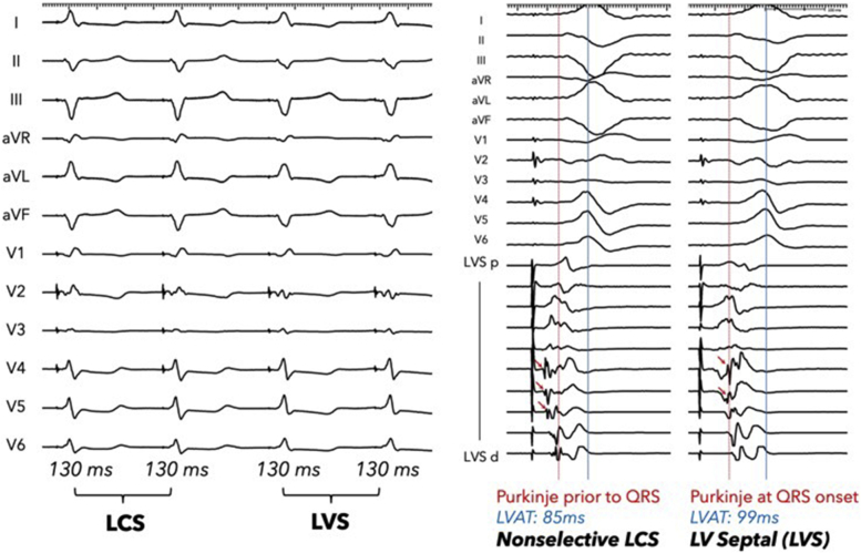
Subtle differences in surface QRS morphology with LCS and LVS. Both exhibit an isoelectric segment after the stimulus; however, intracardiac recordings show evidence of septal capture. High-density mapping of the septum shows presystolic recruitment of Purkinje, which proves LCS, whereas passive Purkinje is still activated at QRS onset during LVS. No data suggest whether these responses are equivalent. LCS = left conduction system; LVAT = left ventricular activation time; LVS = left ventricular (LV) septum.
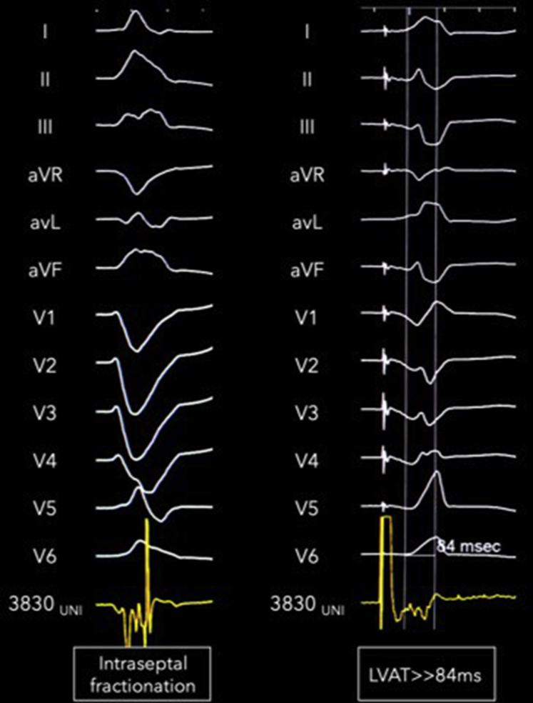
Evidence of intraseptal substrate that impedes the ability to fix the lead deeper and correct wide QRS. Unipolar electrogram shows significant fractionated local recording within the septum. Without including the S-QRS, the left ventricular activation time (LVAT) is already 84 ms from intrinsicoid deflection, signifying inability to achieve cardiac resynchronization therapy by left bundle branch area pacing.
Similar articles
-
Left Bundle Branch Pacing: JACC Review Topic of the Week.J Am Coll Cardiol. 2019 Dec 17;74(24):3039-3049. doi: 10.1016/j.jacc.2019.10.039. Epub 2019 Dec 9. J Am Coll Cardiol. 2019. PMID: 31865972 Review.
-
Permanent His bundle pacing to replace biventricular pacing for cardiac resynchronization therapy.Med Hypotheses. 2017 Nov;109:77-79. doi: 10.1016/j.mehy.2017.09.026. Epub 2017 Sep 28. Med Hypotheses. 2017. PMID: 29150300
-
His bundle pacing after failure of cardiac resynchronization therapy: a case study.J Int Med Res. 2020 May;48(5):300060520923495. doi: 10.1177/0300060520923495. J Int Med Res. 2020. PMID: 32420781 Free PMC article.
-
Permanent His-bundle pacing as an alternative to biventricular pacing for cardiac resynchronization therapy: A multicenter experience.Heart Rhythm. 2018 Mar;15(3):413-420. doi: 10.1016/j.hrthm.2017.10.014. Epub 2017 Oct 12. Heart Rhythm. 2018. PMID: 29031929
-
His Bundle Pacing for Cardiac Resynchronization.Card Electrophysiol Clin. 2018 Sep;10(3):511-517. doi: 10.1016/j.ccep.2018.05.010. Epub 2018 Jul 21. Card Electrophysiol Clin. 2018. PMID: 30172288 Review.
Cited by
-
Ultra-High-Frequency ECG in Cardiac Pacing and Cardiac Resynchronization Therapy: From Technical Concept to Clinical Application.J Cardiovasc Dev Dis. 2024 Feb 23;11(3):76. doi: 10.3390/jcdd11030076. J Cardiovasc Dev Dis. 2024. PMID: 38535099 Free PMC article. Review.
-
Comparative analysis of left bundle branch area pacing in patients with and without a history of open-heart surgery.J Arrhythm. 2025 Jan 30;41(1):e70010. doi: 10.1002/joa3.70010. eCollection 2025 Feb. J Arrhythm. 2025. PMID: 39886037 Free PMC article.
-
State of the Journal 2021: Heart Rhythm O 2.Heart Rhythm O2. 2021 Feb 19;2(1):1-2. doi: 10.1016/j.hroo.2021.01.003. eCollection 2021 Feb. Heart Rhythm O2. 2021. PMID: 34113897 Free PMC article. No abstract available.
-
Gender Differences for His Bundle Pacing Long-Term Performance in the Elderly Population.J Cardiovasc Dev Dis. 2025 Feb 26;12(3):88. doi: 10.3390/jcdd12030088. J Cardiovasc Dev Dis. 2025. PMID: 40137086 Free PMC article.
-
Left bundle branch pacing with and without anodal capture: impact on ventricular activation pattern and acute haemodynamics.Europace. 2023 Oct 5;25(10):euad264. doi: 10.1093/europace/euad264. Europace. 2023. PMID: 37815462 Free PMC article.
References
-
- Durrer D., van Dam R.T., Freud G.E., Janse M.J., Meijler F.L., Arzbaecher R.C. Total excitation of the isolated human heart. Circulation. 1970;41:899–912. - PubMed
-
- Huang W., Su L., Wu S. A novel pacing strategy with low and stable output: pacing the left bundle branch immediately beyond the conduction block. Can J Cardiol. 2017;33:1736 e1731–1736 e1733. - PubMed
-
- Zhang S., Zhou X., Gold M.R. Left bundle branch pacing: JACC review topic of the week. J Am Coll Cardiol. 2019;74:3039–3049. - PubMed
LinkOut - more resources
Full Text Sources
