. 2021 Jul 16;27(40):10394-10404.
doi: 10.1002/chem.202100961. Epub 2021 Jun 10.

Managing Experimental 3D Structures in the Beyond-Rule-of-5 Chemical Space: The Case of Rifampicin

Affiliations

Managing Experimental 3D Structures in the Beyond-Rule-of-5 Chemical Space: The Case of Rifampicin

Giuseppe Ermondi et al. Chemistry. .

Abstract

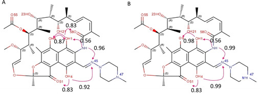
The beyond-Rule-of-5 (bRo5) chemical space is a source of new oral drugs and includes large and flexible compounds. Because of their size and conformational variability, bRo5 molecules assume different privileged conformations in the compartments of human body, i. e., they can exhibit chameleonic properties. The elucidation of the ensemble of 3D structures explored by such molecules under different conditions is therefore critical to check the role played by chameleonicity to modulate cell permeability. Here we characterized the conformational ensembles of rifampicin, a bRo5 drug, in polar and nonpolar solvents and in the solid state. We performed NMR experiments, analyzed their results with a novel algorithm and set-up a pool of ad hoc in silico strategies to investigate crystallographic structures retrieved from the CSD. Moreover, a polarity descriptor often related to permeability (SA-3D-PSA) was calculated for all the conformers and its variation with the environment analyzed. Results showed that the conformational behavior of rifampicin in solution and in the solid state is not superposable. The identification of dynamic intramolecular hydrogen bonds can be assessed by NMR spectroscopy but not by X-ray structures. Moreover, SA-3D-PSA revealed that dynamic IMHBs do not provide rifampicin with chameleonic properties. Overall, this study highlights that the peculiarity of rifampicin, which is cell permeable probably because of the presence of static IMHBs but is devoid of any chameleonic behavior, can be assessed by a proper analysis of experimental 3D structures.

Keywords: NMR spectroscopy; bRo5; chameleonicity; intramolecular hydrogen bonds; macrocycles.


Conflict of interest statement

The authors declare no conflict of interest.

Figures

Figure 1
Figure 1
Rifampicin structure, numbering and ionization properties. A) the three main moieties: the naphthohydroquinone system fused with the furanone ring; the flexible chain (called ansa); the extra‐cycle moiety formed by a (4‐methyl‐1‐piperazinyl)‐iminomethyl group; B) atomic numbering; and C) aqueous ionization profile.
Figure 2
Figure 2
Rifampicin HBP propensity calculated by Mercury: A) neutral and B) zwitterionic form.
Figure 3
Figure 3
Superposition of the X‐ray structures using the shared naphthohydroquinone moiety as a template, the clusters are also shown separately in the same orientation to gain clarity and are colored according to Table 2; A) all structures; B) cluster with YELYAS, YELYIA, YELYOG, YELZAT, YELZEX, YELZIB, YELZOH, YELZUN, YEMBAW, in grey; C) cluster with HAXWUA, MAPHIW, YELYUM, YEMCIF, in red; D) cluster in with YELXUL and YELYEW, in green; E) MAPHES.
Figure 4
Figure 4
Superposition of the representative structures of the four clusters (HAXWUA in red, MAPHES in magenta, YELXUL in green and YELZOH in grey). using different section of the molecules: A) the ansa; B) the (4‐methyl‐1‐piperazinyl)‐iminomethyl group; C) the naphthohydroquinone system.
Figure 5
Figure 5
IMHB pattern deduced from crystal structures: A) zwitterionic form (15 structures); B) neutral form (1 structure). In red IMHBfrac values greater than 0.8, in yellow IMHBfrac values greater than 0.5 and less than 0.8 and, finally, in cyan IMHBfrac values lower than 0.5.
Figure 6
Figure 6
The results of the simulation of neutral rifampicin in chloroform. (a) The time course of the RMSD between the simulated molecule and the minimum‐energy structure indicate conformational changes. (b) The distribution of the dihedral between atoms 12‐52‐29‐28 of the macrocycle, calculated from the simulation. (c) The two peaks in the distribution define two clusters of conformations with different orientations of the macrocycle, labelled as A and B, (d) A clustering analysis of the simulated conformation reveals that clusters A and B can be further partitioned into sub‐clusters with different patterns of hydrogen bonds. (e) The comparison between sub‐clusters A1 and A2 and sub‐clusters B1 and B2 highlights a different orientation of the piperazine ring. (f) The formation probability of hydrogen bonds along the diagonal and their Pearson's correlation coefficients off‐diagonal; all pairs of hydrogen bonds that are able to form are considered.
Figure 7
Figure 7
The results of the simulation of zwitterionic rifampicin in D2O. (a) The time course of the RMSD to the crystallographic structure shows fluctuations between at least three conformations. (b) The distribution associated with two dihedrals of the macrocycles of the three clusters. (d) The result of the clustering analysis. (e) The comparison between sub‐clusters. (f) the formation probability (on the diagonal) and the correlation (off‐diagonal) between hydrogen bonds; all pairs of hydrogen bonds that are able to form are considered.
Figure 8
Figure 8
Superposition of NMR spectroscopy with the crystallographic relevant structures, one for each cluster. Crystallographic structures are colored according to Table 2 whereas different nuances of blue and yellow are used for NMR structure obtained in water and in chloroform, respectively. A) View perpendicular to the naphthohydroquinone moiety; B) lateral view.
Figure 9
Figure 9
SA‐3D‐PSA values calculated on CSD and NMR conformations. A) Bars due to the crystallographic structures are colored on the basis of CSD clusters (Table 2) whereas yellow and orange colors were used for NMR conformations present in chloroform and water, respectively. B) Box plot of SA‐3D‐PSA values calculated on CSD (in cyan and in orange the zwitterionic and the neutral form respectively) and on NMR conformations (in blue and in yellow the zwitterionic and the neutral form respectively, different nuances of blue and yellow are used to distinguish the different clusters).

References

    1. Doak B. C., Over B., Giordanetto F., Kihlberg J., Chem. Biol. 2014, 21, 1115–42. - PubMed
    1. Poongavanam V., Doak B. C., Kihlberg J., Curr. Opin. Chem. Biol. 2018, 44, 23–29. - PubMed
    1. Poongavanam V., Danelius E., Peintner S., Alcaraz L., Caron G., Cummings M. D., Wlodek S., Erdelyi M., Hawkins P. C. D., Ermondi G., Kihlberg J., ACS Omega 2018, 3, 11742–11757. - PMC - PubMed
    1. Ermondi G., Vallaro M., Goetz G. H., Shalaeva M., Caron G., Eur. J. Pharm. Sci. 2020, 146, 105274. - PubMed
    1. Rossi Sebastiano M., Doak B. C. B. C., Backlund M., Poongavanam V., Over B., Ermondi G., Caron G., Matsson P., Kihlberg J., J. Med. Chem. 2018, 61, 4189–4202. - PubMed