

Cytokines: From Clinical Significance to Quantification
- PMID: 34114369
- PMCID: PMC8336501
- DOI: 10.1002/advs.202004433
Cytokines: From Clinical Significance to Quantification
Abstract
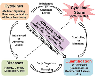
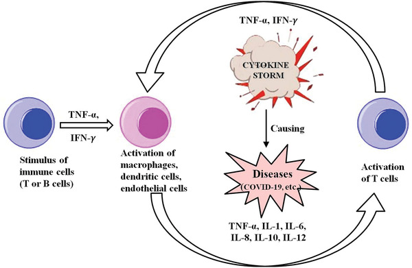
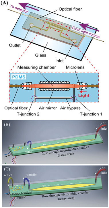
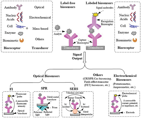
Cytokines are critical mediators that oversee and regulate immune and inflammatory responses via complex networks and serve as biomarkers for many diseases. Quantification of cytokines has significant value in both clinical medicine and biology as the levels provide insights into physiological and pathological processes and can be used to aid diagnosis and treatment. Cytokines and their clinical significance are introduced from the perspective of their pro- and anti-inflammatory effects. Factors affecting cytokines quantification in biological fluids, native levels in different body fluids, sample processing and storage conditions, sensitivity to freeze-thaw, and soluble cytokine receptors are discussed. In addition, recent advances in in vitro and in vivo assays, biosensors based on different signal outputs and intracellular to extracellular protein expression are summarized. Various quantification platforms for high-sensitivity and reliable measurement of cytokines in different scenarios are discussed, and commercially available cytokine assays are compared. A discussion of challenges in the development and advancement of technologies for cytokine quantification that aim to achieve real-time multiplex cytokine analysis for point-of-care situations applicable for both biomedical research and clinical practice are discussed.
Keywords: biosensors; clinical significance; cytokines; in vitro and in vivo assays; quantification.
© 2021 The Authors. Advanced Science published by Wiley-VCH GmbH.
Conflict of interest statement
The authors declare no conflict of interest.
Figures
Similar articles
-
Translational Metabolomics of Head Injury: Exploring Dysfunctional Cerebral Metabolism with Ex Vivo NMR Spectroscopy-Based Metabolite Quantification.In: Kobeissy FH, editor. Brain Neurotrauma: Molecular, Neuropsychological, and Rehabilitation Aspects. Boca Raton (FL): CRC Press/Taylor & Francis; 2015. Chapter 25. In: Kobeissy FH, editor. Brain Neurotrauma: Molecular, Neuropsychological, and Rehabilitation Aspects. Boca Raton (FL): CRC Press/Taylor & Francis; 2015. Chapter 25. PMID: 26269925 Free Books & Documents. Review.
-
Comparison of multiplex platforms for cytokine assessments and their potential use for biomarker profiling in multiple sclerosis.Cytokine. 2017 Mar;91:145-152. doi: 10.1016/j.cyto.2016.12.021. Epub 2017 Jan 9. Cytokine. 2017. PMID: 28082233
-
Point-of-care detection of cytokines in cytokine storm management and beyond: Significance and challenges.View (Beijing). 2021 Aug;2(4):20210003. doi: 10.1002/VIW.20210003. Epub 2021 May 4. View (Beijing). 2021. PMID: 34766163 Free PMC article. Review.
-
Conceptual and methodological issues relevant to cytokine and inflammatory marker measurements in clinical research.Curr Opin Clin Nutr Metab Care. 2010 Sep;13(5):541-7. doi: 10.1097/MCO.0b013e32833cf3bc. Curr Opin Clin Nutr Metab Care. 2010. PMID: 20657280 Free PMC article. Review.
-
A comparison of biofluid cytokine markers across platform technologies: Correspondence or divergence?Cytokine. 2018 Nov;111:481-489. doi: 10.1016/j.cyto.2018.05.032. Epub 2018 Jun 14. Cytokine. 2018. PMID: 29908923 Free PMC article.
Cited by
-
Cytokine Profiling of Plasma and Atherosclerotic Plaques in Patients Undergoing Carotid Endarterectomy.Int J Mol Sci. 2024 Jan 14;25(2):1030. doi: 10.3390/ijms25021030. Int J Mol Sci. 2024. PMID: 38256102 Free PMC article.
-
IL-2 and IL-1β Patient Immune Responses Are Critical Factors in SARS-CoV-2 Infection Outcomes.J Pers Med. 2022 Oct 17;12(10):1729. doi: 10.3390/jpm12101729. J Pers Med. 2022. PMID: 36294868 Free PMC article.
-
Peronema canescens as a Source of Immunomodulatory Agents: A New Opportunity and Perspective.Biology (Basel). 2024 Sep 22;13(9):744. doi: 10.3390/biology13090744. Biology (Basel). 2024. PMID: 39336171 Free PMC article. Review.
-
Anti-inflammatory properties of commonly used psychiatric drugs.Front Neurosci. 2023 Jan 10;16:1039379. doi: 10.3389/fnins.2022.1039379. eCollection 2022. Front Neurosci. 2023. PMID: 36704001 Free PMC article. Review.
-
A Comprehensive Analysis of Cytokine Network in Centenarians.Int J Mol Sci. 2023 Feb 1;24(3):2719. doi: 10.3390/ijms24032719. Int J Mol Sci. 2023. PMID: 36769039 Free PMC article.
References
-
- Kabel A. M., J. Cancer Res. Treat. 2014, 2, 41.
-
- Neurath M. F., Nat. Rev. Immunol. 2014, 14, 329. - PubMed
Publication types
MeSH terms
Substances
Grants and funding
LinkOut - more resources
Full Text Sources
Other Literature Sources