Review
. 2021 Jun 1;14(6):dmm048912.
doi: 10.1242/dmm.048912. Epub 2021 Jun 11.

Mitochondrial function in development and disease

Affiliations
Review

Mitochondrial function in development and disease

Marlies P Rossmann et al. Dis Model Mech. .

Abstract

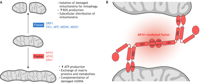
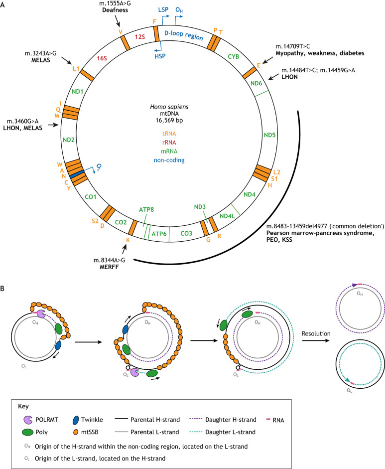
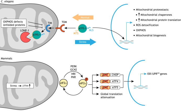
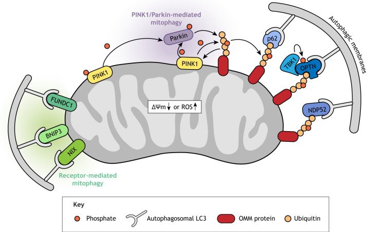
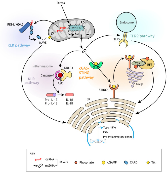
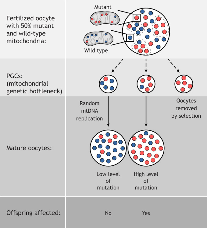
Mitochondria are organelles with vital functions in almost all eukaryotic cells. Often described as the cellular 'powerhouses' due to their essential role in aerobic oxidative phosphorylation, mitochondria perform many other essential functions beyond energy production. As signaling organelles, mitochondria communicate with the nucleus and other organelles to help maintain cellular homeostasis, allow cellular adaptation to diverse stresses, and help steer cell fate decisions during development. Mitochondria have taken center stage in the research of normal and pathological processes, including normal tissue homeostasis and metabolism, neurodegeneration, immunity and infectious diseases. The central role that mitochondria assume within cells is evidenced by the broad impact of mitochondrial diseases, caused by defects in either mitochondrial or nuclear genes encoding for mitochondrial proteins, on different organ systems. In this Review, we will provide the reader with a foundation of the mitochondrial 'hardware', the mitochondrion itself, with its specific dynamics, quality control mechanisms and cross-organelle communication, including its roles as a driver of an innate immune response, all with a focus on development, disease and aging. We will further discuss how mitochondrial DNA is inherited, how its mutation affects cell and organismal fitness, and current therapeutic approaches for mitochondrial diseases in both model organisms and humans.

Keywords: Mitochondrial diseases; Mitochondrial fusion and fission; Mitochondrial unfolded protein response; Mitophagy; mtDNA heteroplasmy and inheritance; mtDNA-mediated innate immune response.


Conflict of interest statement

Competing interests L.I.Z. is a founder of and holds stock in Fate Therapeutics, Camp4 Therapeutics and Scholar Rock, and is a consultant to Celularity. All other authors declare no competing interests.

Figures

Fig. 1.
Fig. 1.
Mitochondrial fusion and fission. (A) Factors involved in these processes and effects on mitochondrial activity. (B) Mitofusin 1 (MFN1)-mediated fusion between two outer mitochondrial membranes. Mitofusins are dynamin-related GTPases essential for mitochondrial fusion, which in turn is crucial for physiological mitochondrial function. Importantly, fusion allows complementation of damaged mtDNA (Nakada et al., 2001). Fusion defects cause neurologic disease (see Table 1). MFN1 is comprised of an N-terminal GTPase domain and two coiled-coil heptad-repeat regions (HR1 and HR2) that are separated by two adjacent small transmembrane domains. This model is based on crystal structures of a truncated version of MFN1 lacking the C-terminal part of the HR1 domain, the transmembrane domain (TM) and the N-terminal part of the HR2 domain (see Cao et al., 2017; Qi et al., 2016). ATP, adenosine triphosphate; GTPase, guanosine triphosphate hydrolysis domain; HD1, helical domain 1, HD2, helical domain 2; mtDNA, mitochondrial DNA; ROS, reactive oxygen species.
Fig. 2.
Fig. 2.
Organization and replication of the human mitochondrial genome. (A) Map of mtDNA. The black arrows point to the nucleotides affected in the indicated diseases; the black bar depicts the span of the ‘common deletion’ in mtDNA, which accounts for a third of Kearns-Sayre syndrome (KSS) cases (see Table 3). HSP, H-strand promoter; LHON, Leber hereditary optic neuropathy; LSP, L-strand promoter; MELAS, Mitochondrial myopathy, encephalopathy, lactic acidosis and stroke-like episodes; MERFF, myoclonic epilepsy with ragged red fibers; NCR, non-coding control region (also known as D-loop region); PEO, progressive external ophthalmoplegia. (B) Strand displacement model of mtDNA replication. After initiation of replication at OH by an RNA primer transcribed by mitochondrial DNA-directed RNA polymerase (POLRMT), POLRMT is replaced by Polγ, which synthesizes the full-length nascent daughter heavy (H)-strand using the light (L)-strand DNA as the template, with the Twinkle helicase moving on the parental H-strand ahead of Polγ and mitochondrial single-stranded DNA (ssDNA)-binding protein (mtSSB) coating the displaced parental H-strand. Once Twinkle reveals OL, a stem-loop forms in the ssDNA of the parental H-strand, allowing the synthesis of a short RNA primer by POLRMT that is used to initiate synthesis of the daughter L-strand by Polγ using the displaced parental H-strand as a template. Twinkle is not required for L-strand synthesis because its template, the displaced H-strand, is unwound and coated with mtSSB. Primer removal and resolution of the hemicatenanes produces two double-stranded daughter mtDNA molecules.
Fig. 3.
Fig. 3.
Models of UPRmt pathways in C. elegans and mammals. In C. elegans, the mitochondrial unfolded protein response (UPRmt) is regulated by the subcellular localization of the transcription factor ATFS-1, which harbors both a mitochondrial targeting sequence (MTS) and a nuclear localization signal (NLS). ATFS-1 is normally efficiently imported into mitochondria through the TOM-TIM mitochondrial translocation complexes and degraded by the protease LONP-1. If ATFS-1 cannot be imported due to mitochondrial stress, ATFS-1 translocates, via the NLS, into the nucleus to activate a broad transcriptional stress response. In mammals, no direct homolog of ATFS-1 has been identified. Rather, the integrated stress response (ISR) is activated via the translation initiation factor eIF2α. Under mitochondrial stress conditions, eIF2α is phosphorylated by four different kinases (PERK, GCN2, HRI or PKR) that are activated by different stimuli. This leads to global translational attenuation, and, at the same time, translation of the transcription factors CHOP, ATF4 and ATF5. This occurs due to skipped translation of the upstream open reading frames (uORFs) that normally inhibit translation of the downstream CHOP, ATF4 and ATF5 coding sequences. OXPHOS, oxidative phosphorylation; ΔΨm, membrane potential.
Fig. 4.
Fig. 4.
Model of ubiquitin-dependent and -independent mitophagy pathways. Mitochondrial stress stabilizes PINK1 on the outer mitochondrial membrane (OMM). PINK1 is activated by autophosphorylation and then phosphorylates Parkin and ubiquitin, both of which activate Parkin's E3 ligase activity. Parkin ubiquitinates several OMM proteins, and the resulting poly-ubiquitin chains in turn serve as additional phosphorylation targets for PINK1, creating a feed-forward loop. The phosphorylated poly-ubiquitin chains trigger the recruitment of the ubiquitin-binding adaptor proteins OPTN, NDP52 and p62, which initiate autophagosome formation by directly binding to the autophagosomal light chain 3 (LC3) protein through their LC-interacting region motifs. OPTN's affinity for ubiquitin chains is enhanced by its phosphorylation, and TANK binding kinase 1 (TBK1). Receptor-mediated mitophagy relies on various OMM proteins including BNIP3, NIX and FUNDC1, which directly interact with LC3 to mediate autophagosome formation.
Fig. 5.
Fig. 5.
Innate immune response pathways elicited by damage-associated molecular patterns (DAMPs). Mitochondrial stress or Bak/Bax-driven OMM permeabilization can lead to release of mtDNA or dsRNA into the cytosol, triggering a cascade that results in activated expression of type I interferon and pro-inflammatory cytokine genes. Cytosolic mtDNA can bind the DNA-sensing protein cGAS that catalyzes the production of 2′3′-cyclic GMP-AMP (cGAMP), which in turn binds the adaptor molecule STING1 on the ER, activating TBK1. TBK1 phosphorylates and thus induces the translocation of the transcription factor IRF3 into the nucleus, where it activates type I interferon genes. mtDNA can also trigger a pro-inflammatory or type I interferon response via binding to Toll-like receptor 9 (TLR9) located on endosomes. In addition, mtDNA can be an endogenous agonist of cytosolic inflammasomes, multi-subunit complexes consisting of the receptor NLRP3 (or NLRC4, or AIM2), the adaptor ASC and the inflammatory cysteine protease caspase-1, which processes pro-IL-1β and pro-IL-18 into their mature forms. Double-stranded RNA (dsRNA) is recognized by the retinoic acid-inducible gene-I-like receptors (RLRs) RIG-I or melanoma differentiation-associated gene 5 (MDA5), which bind to mitochondrial antiviral signaling protein (MAVS) through homotypic caspase activation and recruitment domain (CARD)-CARD interactions. MAVS then recruits various molecules to transduce a downstream signal to the nucleus, resulting in the activation of target genes. ER, endoplasmic reticulum; IFN, interferon; ISG, interferon-stimulatory gene; mtROS, mitochondrial reactive oxygen species.
Fig. 6.
Fig. 6.
The mitochondrial bottleneck in the female germline and its consequences for the offspring. The penetrance of phenotypes depends on the level of heteroplasmy, which represents mutant mtDNA load. A threshold of 65-90% mutant mtDNA needs to be surpassed for a respiratory chain deficiency to manifest. PGC, primordial germ cell.

References

    1. Abudayyeh, O. O., Gootenberg, J. S., Konermann, S., Joung, J., Slaymaker, I. M., Cox, D. B., Shmakov, S., Makarova, K. S., Semenova, E., Minakhin, L., et al. (2016). C2c2 is a single-component programmable RNA-guided RNA-targeting CRISPR effector. Science 353, aaf5573. 10.1126/science.aaf5573 - DOI - PMC - PubMed
    1. Acin-Perez, R., Bayona-Bafaluy, M. P., Bueno, M., Machicado, C., Fernandez-Silva, P., Perez-Martos, A., Montoya, J., Lopez-Perez, M. J., Sancho, J. and Enriquez, J. A. (2003). An intragenic suppressor in the cytochrome c oxidase I gene of mouse mitochondrial DNA. Hum. Mol. Genet. 12, 329-339. 10.1093/hmg/ddg021 - DOI - PubMed
    1. Aguirre, S., Luthra, P., Sanchez-Aparicio, M. T., Maestre, A. M., Patel, J., Lamothe, F., Fredericks, A. C., Tripathi, S., Zhu, T., Pintado-Silva, J., et al. (2017). Dengue virus NS2B protein targets cGAS for degradation and prevents mitochondrial DNA sensing during infection. Nat Microbiol 2, 17037. 10.1038/nmicrobiol.2017.37 - DOI - PMC - PubMed
    1. Ahlqvist, K. J., Hämäläinen, R. H., Yatsuga, S., Uutela, M., Terzioglu, M., Götz, A., Forsström, S., Salven, P., Angers-Loustau, A., Kopra, O. H., et al. (2012). Somatic progenitor cell vulnerability to mitochondrial DNA mutagenesis underlies progeroid phenotypes in Polg mutator mice. Cell Metab. 15, 100-109. 10.1016/j.cmet.2011.11.012 - DOI - PubMed
    1. Ahlqvist, K. J., Leoncini, S., Pecorelli, A., Wortmann, S. B., Ahola, S., Forsstrom, S., Guerranti, R., De Felice, C., Smeitink, J., Ciccoli, L., et al. (2015). MtDNA mutagenesis impairs elimination of mitochondria during erythroid maturation leading to enhanced erythrocyte destruction. Nat. Commun. 6, 6494. 10.1038/ncomms7494 - DOI - PubMed

Publication types