. 2021 Jul 1;321(1):H185-H196.
doi: 10.1152/ajpheart.00118.2021. Epub 2021 Jun 11.

Apigenin restores endothelial function by ameliorating oxidative stress, reverses aortic stiffening, and mitigates vascular inflammation with aging

Affiliations

Apigenin restores endothelial function by ameliorating oxidative stress, reverses aortic stiffening, and mitigates vascular inflammation with aging

Zachary S Clayton et al. Am J Physiol Heart Circ Physiol. .

Abstract

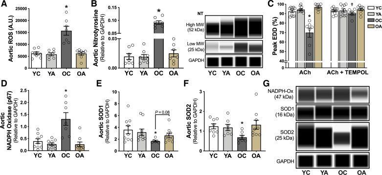
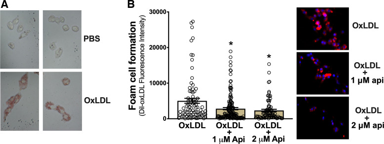
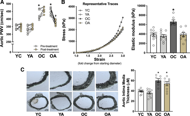
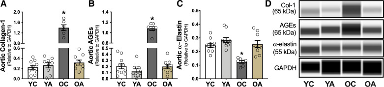
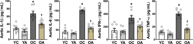
We assessed the efficacy of oral supplementation with the flavanoid apigenin on arterial function during aging and identified critical mechanisms of action. Young (6 mo) and old (27 mo) C57BL/6N mice (model of arterial aging) consumed drinking water containing vehicle (0.2% carboxymethylcellulose; 10 young and 7 old) or apigenin (0.5 mg/mL in vehicle; 10 young and 9 old) for 6 wk. In vehicle-treated animals, isolated carotid artery endothelium-dependent dilation (EDD), bioassay of endothelial function, was impaired in old versus young (70% ± 9% vs. 92% ± 1%, P < 0.0001) due to reduced nitric oxide (NO) bioavailability. Old mice had greater arterial reactive oxygen species (ROS) production and oxidative stress (higher nitrotyrosine) associated with greater nicotinamide adenine dinucleotide phosphate oxidase (oxidant enzyme) and lower superoxide dismutase 1 and 2 (antioxidant enzymes); ex vivo administration of Tempol (antioxidant) restored EDD to young levels, indicating ROS-mediated suppression of EDD. Old animals also had greater aortic stiffness as indicated by higher aortic pulse wave velocity (PWV, 434 ± 9 vs. 346 ± 5 cm/s, P < 0.0001) due to greater intrinsic aortic wall stiffness associated with lower elastin levels and higher collagen, advanced glycation end products (AGEs), and proinflammatory cytokine abundance. In old mice, apigenin restored EDD (96% ± 2%) by increasing NO bioavailability, normalized arterial ROS, oxidative stress, and antioxidant expression, and abolished ROS inhibition of EDD. Moreover, apigenin prevented foam cell formation in vitro (initiating step in atherosclerosis) and mitigated age-associated aortic stiffening (PWV 373 ± 5 cm/s) by normalizing aortic intrinsic wall stiffness, collagen, elastin, AGEs, and inflammation. Thus, apigenin is a promising therapeutic for arterial aging.NEW & NOTEWORTHY Our study provides novel evidence that oral apigenin supplementation can reverse two clinically important indicators of arterial dysfunction with age, namely, vascular endothelial dysfunction and large elastic artery stiffening, and prevents foam cell formation in an established cell culture model of early atherosclerosis. Importantly, our results provide extensive insight into the biological mechanisms of apigenin action, including increased nitric oxide bioavailability, normalization of age-related increases in arterial ROS production and oxidative stress, reversal of age-associated aortic intrinsic mechanical wall stiffening and adverse remodeling of the extracellular matrix, and suppression of vascular inflammation. Given that apigenin is commercially available as a dietary supplement in humans, these preclinical findings provide the experimental basis for future translational studies assessing the potential of apigenin to treat arterial dysfunction and reduce cardiovascular disease risk with aging.

Keywords: aortic pulse wave velocity; atherosclerosis; endothelium-dependent dilation; nutraceutical; reactive oxygen species.


Conflict of interest statement

No conflicts of interest, financial or otherwise, are declared by the authors.

Figures

Figure 1.
Figure 1.
Apigenin selectively enhances endothelial function in old mice by increasing nitric oxide (NO) bioavailability. A: carotid artery dose responses to the endothelium-dependent dilator acetylcholine (ACh) in young and old controls (YC and OC) and young and old apigenin-supplemented (YA and OA) mice (n = 7–10/group) with and without coadministration of the NO synthase inhibitor NG-nitro-l-arginine methyl ester (l-NAME). B: NO-mediated dilation [peak dilation of ACh (−) peak dilation of ACh + l-NAME] (n = 7–10/group). C: dose responses to the endothelium-independent dilator sodium nitroprusside (SNP) (n = 7–10/group). Values are means ± SE; n, number of mice/group. *P < 0.05 vs. all other groups (under ACh-only conditions in A).
Figure 2.
Figure 2.
Apigenin supplementation ameliorates age-related vascular oxidative stress. A: aortic reactive oxygen species (ROS) production [electron paramagnetic resonance spectroscopy absorbance units (AU)] in young and old controls (YC and OC) and young and old apigenin-supplemented (YA and OA) mice (n = 7–10/group). B: aortic nitrotyrosine (NT) abundance and representative images in YC, YA, OC, and OA mice (n = 7–10/group). Data are the sum of the high and low molecular weight bands normalized to glyceraldehyde 3-phosphate dehydrogenase (GAPDH). C: maximal carotid artery dose response to the endothelium-dependent dilator acetylcholine (ACh) in YC, YA, OC, and OA mice in the presence or absence of the ROS scavenger Tempol (n = 7–10/group). Aortic abundance and representative images of NADPH oxidase (D and G); superoxide dismutase (SOD) 1 (E and G); and SOD2 (F and G), all of which are normalized to GAPDH. Values are means ± SE; n, number of mice/group. *P < 0.05 vs. all other groups; †P < 0.05 ACh vs. ACh + Tempol. EDD, endothelium-dependent dilation; MW, molecular weight; NADPH-ox, nicotinamide adenine dinucleotide phosphate oxidase.
Figure 3.
Figure 3.
Apigenin prevents oxidized low-density lipoprotein (oxLDL)-induced foam cell formation in cultured RAW 264.7 macrophages. A: qualitative proof of concept images that oxLDL stimulates foam cell formation as defined by greater Oil Red O uptake in Raw 264.7 macrophages when exposed to oxLDL [in phosphate-buffered saline (PBS) vs. PBS alone]. B: foam cell formation, quantified as fluorescent DiI-oxLDL incorporation into Raw 264.7 macrophages, following incubation with 0.5 µg/mL DiI-oxLDL + 50 µg/mL oxLDL; 0.5 µg/mL DiI-oxLDL + 50 µg/mL oxLDL + 1 µM apigenin (api); or 0.5 µg/mL DiI-oxLDL + 50 µg/mL oxLDL + 2 µM api. Representative images are provided on the right. Each condition was tested in quadruplicate on two separate days, with n = 8/condition/day. Values are means ± SE; n, number of imaged wells/condition per day. *P < 0.05 vs. oxLDL.
Figure 4.
Figure 4.
Apigenin reverses age-related aortic stiffening. A: aortic pulse wave velocity (PWV) in young and old controls (YC and OC) and young and old apigenin-supplemented (YA and OA) mice (n = 7–10/group). B: representative stress-strain curve of an aorta ring from YC, YA, OC, and OA mice for determination of ex vivo intrinsic mechanical wall stiffness. Aortic elastic modulus (calculated as the slope of the final four points in the stress-strain curve) in YC, YA, OC, and OA mice. C: intima-media thickness of the aorta with representative images of whole aortic sections (bottom) and enlargements of the same sections (top) included to the left of the mean data (n = 7–10/group). Values are means ± SE; n, number of mice/group. *P < 0.05 vs. all other groups (aortic PWV, pretreatment); †P < 0.05 pretreatment vs. posttreatment within group.
Figure 5.
Figure 5.
Apigenin supplementation reverses age-related increases in aortic collagen and advanced glycation end products (AGEs) and prevents the age-related reduction in aortic elastin. A and D: aortic abundance of collagen-1 (Col-1) in young and old controls (YC and OC) and young and old apigenin-supplemented (YA and OA) mice (n = 7–10/group). B and D: aortic abundance of AGEs in YC, YA, OC, and OA mice. C and D: aortic abundance of α-elastin in YC, YA, OC, and OA mice. Data are normalized to glyceraldehyde 3-phosphate dehydrogenase (GAPDH). Values are means ± SE; n, number of mice/group. *P < 0.05 vs. all other groups.
Figure 6.
Figure 6.
Apigenin supplementation attenuates age-related vascular inflammation. Aortic abundance of interleukin-1β (IL-1β), interleukin-6 (IL-6), interferon γ (IFNγ), and tumor necrosis factor α (TNFα) in young and old controls (YC and OC) and young and old apigenin-supplemented (YA and OA) mice (n = 7–10/group). Values are means ± SE; n, number of mice/group. *P < 0.05 vs. all other groups; †P < 0.05 vs. all other groups.

References

    1. Benjamin EJ, Virani SS, Callaway CW, Chamberlain AM, Chang AR, Cheng S. Heart disease and stroke statistics-2018 update: a report from the American Heart Association. Circulation 137: e67–e492, 2018. doi:10.1161/CIR.0000000000000558. - DOI - PubMed
    1. Lakatta EG, Levy D. Arterial and cardiac aging: major shareholders in cardiovascular disease enterprises: Part I: aging arteries: a “set up” for vascular disease. Circulation 107: 139–146, 2003. doi:10.1161/01.cir.0000048892.83521.58. - DOI - PubMed
    1. Brunt VE, Gioscia-Ryan RA, Richey JJ, Zigler MC, Cuevas LM, Gonzalez A, Vázquez-Baeza Y, Battson ML, Smithson AT, Gilley AD, Ackermann G, Neilson AP, Weir T, Davy KP, Knight R, Seals DR. Suppression of the gut microbiome ameliorates age-related arterial dysfunction and oxidative stress in mice. J Physiol 597: 2361–2378, 2019. doi:10.1113/JP277336. - DOI - PMC - PubMed
    1. Seals DR, Jablonski KL, Donato AJ. Aging and vascular endothelial function in humans. Clin Sci (Lond) 120: 357–375, 2011. doi:10.1042/CS20100476. - DOI - PMC - PubMed
    1. El Assar M, Angulo J, Vallejo S, Peiró C, Sánchez-Ferrer CF, Rodríguez-Mañas L. . Mechanisms involved in the aging-induced vascular dysfunction. Front Physiol 3: 132, 2012. doi:10.3389/fphys.2012.00132. - DOI - PMC - PubMed

Publication types