. 2021 Dec 16;144(11):3461-3476.
doi: 10.1093/brain/awab217.

TDP-43 stabilizes G3BP1 mRNA: relevance to amyotrophic lateral sclerosis/frontotemporal dementia

Affiliations

TDP-43 stabilizes G3BP1 mRNA: relevance to amyotrophic lateral sclerosis/frontotemporal dementia

Hadjara Sidibé et al. Brain. .

Abstract

TDP-43 nuclear depletion and concurrent cytoplasmic accumulation in vulnerable neurons is a hallmark feature of progressive neurodegenerative proteinopathies such as amyotrophic lateral sclerosis (ALS) and frontotemporal dementia (FTD). Cellular stress signalling and stress granule dynamics are now recognized to play a role in ALS/FTD pathogenesis. Defective stress granule assembly is associated with increased cellular vulnerability and death. Ras-GAP SH3-domain-binding protein 1 (G3BP1) is a critical stress granule assembly factor. Here, we define that TDP-43 stabilizes G3BP1 transcripts via direct binding of a highly conserved cis regulatory element within the 3' untranslated region. Moreover, we show in vitro and in vivo that nuclear TDP-43 depletion is sufficient to reduce G3BP1 protein levels. Finally, we establish that G3BP1 transcripts are reduced in ALS/FTD patient neurons bearing TDP-43 cytoplasmic inclusions/nuclear depletion. Thus, our data indicate that, in ALS/FTD, there is a compromised stress granule response in disease-affected neurons due to impaired G3BP1 mRNA stability caused by TDP-43 nuclear depletion. These data implicate TDP-43 and G3BP1 loss of function as contributors to disease.

Keywords: G3BP1; TDP-43; amyotrophic lateral sclerosis; frontotemporal dementia; stress granules.


Figures

Figure 1
Figure 1
TDP-43 nuclear depletion and ALS-related species induce G3BP1 downregulation. (A) HeLa cells were transfected with siTDP-43 or siControl (siCTL), then transfected with empty plasmid (Mock) or Flag-TDP-43ΔNLS and immunoblotted. (B) Quantification via densitometry of G3BP1 protein levels normalized to actin. (C) Protein from SH-SY5Y wild-type and TDP-43N352S/N352S cells were extracted, and immunoblotted. Quantification via densitometry of (D) TDP-43 and (E) G3BP1 protein levels normalized to actin. (F) HeLa cells were transfected with empty plasmid (Mock) or Flag-tagged TDP-35 and immunoblotted. Quantification via densitometry of (G) TDP-43 and (H) G3BP1 protein levels normalized to actin. Data from three to five independent experiments are expressed as the mean fold change ± SEM; Unpaired t-test *P < 0.05; **P < 0.01. WT = wild-type.
Figure 2
Figure 2
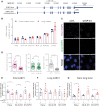
TDP-43 depletion only affects the short G3BP1 transcript. (A) Schematic of Genome Reference Consortium Human GRCh38.p12 G3BP1 transcripts. Long transcript has 8901-nt 3ʹ UTR while the shorter version has a 1466-nt 3ʹ UTR. (B) RT-qPCR for total G3BP1, long G3BP1 transcripts and GAPDH of extracted RNA from HeLa, HEK293, SK-N-SH, SH-SY5Y, U2OS and Mo3.13 cells, normalized with GAPDH and 18S, n = 3–4. (C) Representative images of smFISH in HeLa cells treated with siControl and siTDP-43, using probes against total G3BP1 (magenta) and long G3BP1 (green) mRNA. Cell nuclei were counterstained with Hoechst. Yellow foci show co-localization of total and long signal. (D) Total G3BP1 and long G3BP1 were quantified and short G3BP1 was calculated as the number of long foci subtracted from total magenta foci for each cell. Data are expressed as the mean ± SEM, n = 3, N = 45. Unpaired t-test, ****P < 0.0001, scale bar = 25 μm. (E and F) RT-qPCR for total G3BP1 and long G3BP1 transcripts extracted RNA from HeLa, SK-N-SH and SH-SY5Y cells transfected with siControl or siTDP-43 and normalized with GAPDH and 18S. (G) Ratio of the levels of long G3BP1 transcripts:total G3BP1, mean ± SEM, n = 3–5, unpaired t-test *P < 0.05, **P < 0.01.
Figure 3
Figure 3
TDP-43 regulates and stabilizes G3BP1 via its 3ʹ UTR. (A) HeLa cells were transfected with siTDP-43 or siCTL, then cotransfected with the indicated reporter plasmids. Luciferase activity of G3BP1 3ʹ UTR is expressed relative to siCTL cells. GADPH and R03 (random sequences) are used as controls, mean ± SEM, n = 4, unpaired t-test **P < 0.01, ***P < 0.001. (B) RT-qPCR for His-G3BP1 transcripts, normalized to 18S following doxycycline treatment for 6 h to shut off expression, n = 3. (C) Half-lives of His-G3BP1 transcripts in HeLa-Tet-off cells transfected with siCTL compared to cells transfected with siTDP-43, n = 3, unpaired t-test **P < 0.01.
Figure 4
Figure 4
Identification of a conserved element in the G3BP1 3ʹ UTR. (A) Schematic representation of the conserved element identified in the human G3BP1 3ʹ UTR between nt 319 and 372 (GRCh38/hg38 chr5:151 183 766–151 183 816). The blue lines represent TDP-43 binding sites suggested by RNAmap. (B) Consensus sequence of the conserved sequence obtained from 13 species using the consensus sequence generator MEME. (C) RT-qPCR for gtbp-1 of extracted RNA from N2 (wild-type control) and tdp-1(ok803) null worms. n = 3, unpaired t-test *P < 0.05. (D) Schematic representation of the G3BP1 3ʹ UTR constructs used. The red rectangle represents the conserved element. (E) HeLa cells were transfected with siTDP-43 or siCTL, then cotransfected with the indicated reporter plasmids. GADPH and R03 (random sequences) are negative controls and HDAC6 is a positive control, mean ± SEM, n = 3–8, unpaired t-test *P < 0.05, **P < 0.01. (F) RT-qPCR for His-G3BP1Δ341-357 transcript, normalized to 18S following doxycycline treatment for 6 h to shut off expression. (G) Half-life of His-G3BP1Δ341-357 transcript in HeLa-Tet-off cells transfected with siCTL compared to cells transfected with siTDP-43, n = 3.
Figure 5
Figure 5
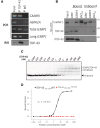
TDP-43 directly binds the conserved element. (A) TDP-43 protein and its associated transcripts were co-immunoprecipitated from HeLa cells homogenates. mRNAs binding to TDP-43 were extracted, reverse transcribed and amplified for G3BP1 transcripts, CaMKII (positive control) and HSPA1A (negative control), showing that TDP-43 binds total G3BP1 transcripts and the longer transcripts. Flag-immunoprecipitation serves as a control to demonstrate specificity of TDP-43 immunoprecipitation. (B) Proteins from whole HeLa cell extracts or TDP-43 recombinant protein (rTDP-43) were pulled down using biotinylated RNA probes containing the TDP-43 binding sequence in GRN (positive control), G3BP1 3ʹ UTRnt319-372, or (AC)12 (negative control). For AC-repeat binding, hnRNP L served as a positive control. (C) Typical EMSA performed with 10 pM 5′-32P-labelled RNA and increasing concentrations of TDP-43102-269. (D) Typical binding curve using wild-type TDP-43102-269 (black line) and the F147L/F149L mutant of TDP-43102-269 (red line). IP = immunoprecipitation; WB = western blot; WCE = whole-cell extract; WT = wild-type.
Figure 6
Figure 6
Changes in TDP-43 and G3BP1 expression in injured motor neurons following medial axotomy. (A) Schematic of the medial site of axotomy. (B) NBA score of the axotomized mice at Days 1, 3, 5 and 7. Data are expressed as the mean ± SEM; Unpaired t-test ****P < 0.0001. (C) Representative images of control non-axotomized (ipsilateral) and axotomized mice (contralateral) spinal cord tissues at Day 7, showing injured spinal motor neurons of the ventral horn using mouse TDP-43, rabbit G3BP1 and guinea pig FluoroGold antibody. The grey line shows the cell boundary. (D) Fluorescence intensity of nuclear TDP-43 and total G3BP1 for the control non-injured and injured motor neurons at Day 7, n = 3 mice and N = 10 cells per mouse. Data are expressed as the mean ± SEM; two-way ANOVA ****P < 0.0001. Scale bar = 10 µm.
Figure 7
Figure 7
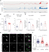
G3BP1 short mRNA is destabilized and reduced in patients with TDP-43-mediated neurodegeneration. (A) Sashimi plots of GEO dataset GSE67196 for human G3BP1 in cerebellum and frontal cortex of control case, showing reduced reads for the long G3BP1 3ʹ UTR in the frontal cortex. (B) Comparison of G3BP1 levels (both transcripts) in human cerebellum and frontal cortex in healthy controls. *P < 0.05. (C) Quantification of polyadenylation site usage in G3BP1 transcripts in lumbar spinal motor neurons isolated by laser capture microdissection (GEO dataset GSE103225). Data are expressed as the mean ± SEM; unpaired t-test ***P < 0.001. (D) RT-qPCR for total G3BP1 and long G3BP1 transcripts extracted RNA from I3 neurons derived from TDP-43M337V and isogenic control fibroblast, mean ± SEM, n = 3, paired t-test *P < 0.05. Normalized to GAPDH and 18S. (E) Neurons in the orbitofrontal cortex of ALS and ALS/FTLD cases showing normal nuclear localization of TDP-43 (top row, green) or nuclear TDP-43 depletion/cytoplasmic TDP-43 accumulation (bottom row, green). G3BP1 mRNA (magenta dots) is labelled with RNAscope probes, nuclei are marked with DAPI (blue). (F) Quantification of G3BP1 mRNA signals in neurons of the orbitofrontal cortex of ALS and ALS/FTLD with or without TDP-43 pathology (defined as obvious cytoplasmic accumulations or reduced nuclear TDP-43 levels). Data are expressed as the mean ± SEM; one-way ANOVA ****P < 0.0001. Scale bar = 10 μm.

References

    1. Taylor JP, Brown RH Jr, Cleveland DW.. Decoding ALS: From genes to mechanism. Nature. 2016;539(7628):197–206. - PMC - PubMed
    1. Kuo PH, Chiang CH, Wang YT, Doudeva LG, Yuan HS.. The crystal structure of TDP-43 RRM1-DNA complex reveals the specific recognition for UG- and TG-rich nucleic acids. Nucleic Acids Res. 2014;42(7):4712–22. - PMC - PubMed
    1. Mompean M, Romano V, Pantoja-Uceda D, et al.The TDP-43 N-terminal domain structure at high resolution. Febs J. 2016;283(7):1242–60. - PubMed
    1. Afroz T, Hock E-M, Ernst P, et al.Functional and dynamic polymerization of the ALS-linked protein TDP-43 antagonizes its pathologic aggregation. Nat Commun. 2017;8(1):45. - PMC - PubMed
    1. Neumann M, Sampathu DM, Kwong LK, et al.Ubiquitinated TDP-43 in frontotemporal lobar degeneration and amyotrophic lateral sclerosis. Science. 2006;314(5796):130–133. - PubMed

Publication types

MeSH terms

Supplementary concepts