

FHOD1 and FMNL1 formin proteins in intestinal gastric cancer: correlation with tumor-infiltrating T lymphocytes and molecular subtypes
- PMID: 34115237
- PMCID: PMC8502136
- DOI: 10.1007/s10120-021-01203-7
FHOD1 and FMNL1 formin proteins in intestinal gastric cancer: correlation with tumor-infiltrating T lymphocytes and molecular subtypes
Abstract
Background: Gastric cancer (GC) is the third most common cause of cancer death. Intestinal type GC is a molecularly diverse disease. Formins control cytoskeletal processes and have been implicated in the progression of many cancers. Their clinical significance in GC remains unclear. Here, we characterize the expression of formin proteins FHOD1 and FMNL1 in intestinal GC tissue samples and investigate their association with clinical parameters, GC molecular subtypes and intratumoral T lymphocytes.
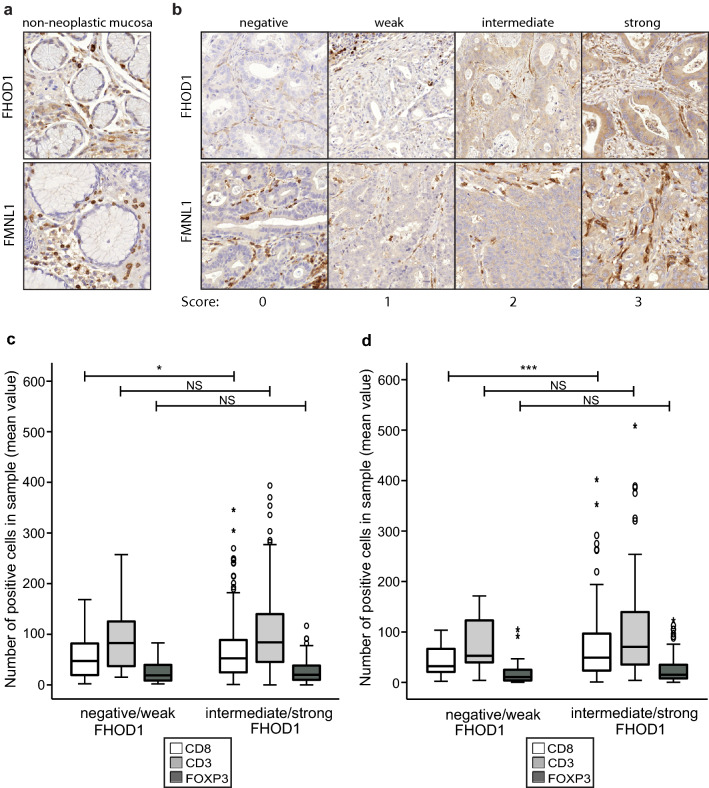
Methods: The prognostic significance of FHOD1 and FMNL1 mRNA expression was studied with Kaplan-Meier analyses in an online database. The expression of FHOD1 and FMNL1 proteins was characterized in GC cells, and in non-neoplastic and malignant tissues utilizing tumor microarrays of intestinal GC representing different molecular subtypes. FHOD1 and FMNL1 expression was correlated with clinical parameters, molecular features and T lymphocyte infiltration. Immunohistochemical expression of neither formin correlated with survival.
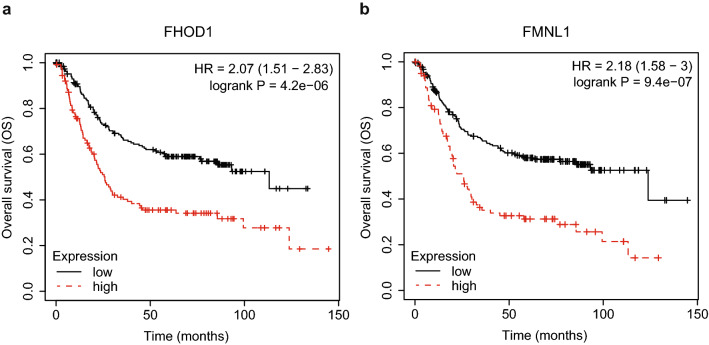
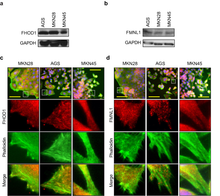
Results: Kaplan-Meier analysis associated high FHOD1 and FMNL1 mRNA expression with reduced overall survival (OS). Characterization of FHOD1 and FMNL1 in GC cells showed cytoplasmic expression along the actin filaments. Similar pattern was recapitulated in GC tissue samples. Elevated FMNL1 was associated with larger tumor size and higher disease stage. Downregulation of FHOD1 associated with TP53-mutated GC tumors. Tumor cell FHOD1 expression strongly correlated with high numbers of tumor-infiltrating CD8 + lymphocytes.
Conclusions: FHOD1 and FMNL1 proteins are expressed in the tumor cells of intestinal GC and significantly associate with clinical parameters without direct prognostic significance. FHOD1 correlates with high intratumoral CD8 + T lymphocyte infiltration in this cohort.
Keywords: FHOD1; FMNL1; Formins; Gastric cancer; T lymphocytes.
© 2021. The Author(s).
Conflict of interest statement
The authors declare that they have no conflict of interests.
Figures
Similar articles
-
Role of FHOD1 in tumor cells and tumor immune microenvironment.Front Immunol. 2025 Apr 29;16:1514488. doi: 10.3389/fimmu.2025.1514488. eCollection 2025. Front Immunol. 2025. PMID: 40364836 Free PMC article. Review.
-
Association of tumor-infiltrating T lymphocytes with intestinal-type gastric cancer molecular subtypes and outcome.Virchows Arch. 2021 Apr;478(4):707-717. doi: 10.1007/s00428-020-02932-3. Epub 2020 Sep 21. Virchows Arch. 2021. PMID: 32954467 Free PMC article.
-
Systematic Characterization of the Expression and Prognostic Values of Formin-Like Gene Family in Gastric Cancer.DNA Cell Biol. 2020 Sep;39(9):1664-1677. doi: 10.1089/dna.2020.5508. Epub 2020 Jun 16. DNA Cell Biol. 2020. PMID: 32551946
-
Multiple formin proteins participate in glioblastoma migration.BMC Cancer. 2020 Jul 29;20(1):710. doi: 10.1186/s12885-020-07211-7. BMC Cancer. 2020. PMID: 32727404 Free PMC article.
-
Formins as effector proteins of Rho GTPases.Small GTPases. 2014;5:e29513. doi: 10.4161/sgtp.29513. Epub 2014 Jun 10. Small GTPases. 2014. PMID: 24914801 Free PMC article. Review.
Cited by
-
Role of FHOD1 in tumor cells and tumor immune microenvironment.Front Immunol. 2025 Apr 29;16:1514488. doi: 10.3389/fimmu.2025.1514488. eCollection 2025. Front Immunol. 2025. PMID: 40364836 Free PMC article. Review.
-
Pan-cancer analysis revealing DAAM1 as a novel predictive biomarker for PD-1/PD-L1 blockade in clear cell renal cell carcinoma.MedComm (2020). 2022 Oct 27;3(4):e177. doi: 10.1002/mco2.177. eCollection 2022 Dec. MedComm (2020). 2022. PMID: 36311172 Free PMC article. No abstract available.
-
The role of Golgi complex proteins in cell division and consequences of their dysregulation.Front Cell Dev Biol. 2025 Jan 7;12:1513472. doi: 10.3389/fcell.2024.1513472. eCollection 2024. Front Cell Dev Biol. 2025. PMID: 39839669 Free PMC article. Review.
-
Formins in Human Disease.Cells. 2021 Sep 27;10(10):2554. doi: 10.3390/cells10102554. Cells. 2021. PMID: 34685534 Free PMC article. Review.
References
MeSH terms
Substances
LinkOut - more resources
Full Text Sources
Medical
Research Materials
Miscellaneous
