. 2021 Aug;38(4):401-410.
doi: 10.1007/s10585-021-10103-0. Epub 2021 Jun 11.

CXCL2-CXCR2 axis mediates αV integrin-dependent peritoneal metastasis of colon cancer cells

Affiliations

CXCL2-CXCR2 axis mediates αV integrin-dependent peritoneal metastasis of colon cancer cells

Mattias Lepsenyi et al. Clin Exp Metastasis. 2021 Aug.

Erratum in

Abstract

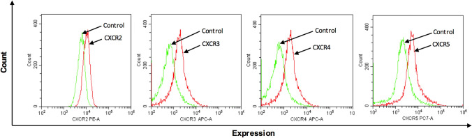
Peritoneal metastasis is an insidious aspect of colorectal cancer. The aim of the present study was to define mechanisms regulating colon cancer cell adhesion and spread to peritoneal wounds after abdominal surgery. Mice was laparotomized and injected intraperitoneally with CT-26 colon carcinoma cells and metastatic noduli in the peritoneal cavity was quantified after treatment with a CXCR2 antagonist or integrin-αV-antibody. CT-26 cells expressed cell surface chemokine receptors CXCR2, CXCR3, CXCR4 and CXCR5. Stimulation with the CXCR2 ligand, CXCL2, dose-dependently increased proliferation and migration of CT-26 cells in vitro. The CXCR2 antagonist, SB225002, dose-dependently decreased CXCL2-induced proliferation and migration of colon cancer cells in vitro. Intraperitoneal administration of CT-26 colon cancer cells resulted in wide-spread growth of metastatic nodules at the peritoneal surface of laparotomized animals. Laparotomy increased gene expression of CXCL2 at the incisional line. Pretreatment with CXCR2 antagonist reduced metastatic nodules by 70%. Moreover, stimulation with CXCL2 increased CT-26 cell adhesion to extracellular matrix (ECM) proteins in a CXCR2-dependent manner. CT-26 cells expressed the αV, β1 and β3 integrin subunits and immunoneutralization of αV abolished CXCL2-triggered adhesion of CT-26 to vitronectin, fibronectin and fibrinogen. Finally, inhibition of the αV integrin significantly attenuated the number of carcinomatosis nodules by 69% in laparotomized mice. These results were validated by use of the human colon cancer cell line HT-29 in vitro. Our data show that colon cancer cell adhesion and growth on peritoneal wound sites is mediated by a CXCL2-CXCR2 signaling axis and αV integrin-dependent adhesion to ECM proteins.

Keywords: Chemokines; Chemotaxis; Integrins; Metastasis; Peritoneal carcinomatosis.


Conflict of interest statement

Authors have no financial conflicts of interests.

Figures

Fig. 1
Fig. 1
Chemokine receptor expression on colon cancer cells. Single-cell suspensions were prepared from confluent CT-26 cells and stained as outlined in Materials and Methods. Unstained cells were used as negative control and single tube staining used for each receptor. n = 4
Fig. 2
Fig. 2
CXCL2 induces CT-26 colon cancer cell proliferation and migration. In vitro cell proliferation and migration in response to 24 h stimulation CXCL2 (10–200 ng/ml) without (A, B) or with (C, D) CXCR2 antagonist (SB225002) (0.2–1 μM). Proliferation was determined fluorometrically after adding calcein AM to cells as described in Materials and Methods. Migration was quantified by counting cells in High Power Fields in 5 different fields. Data represents mean ± SEM and n = 4. #P < 0,05 vs Ctrl and *P < 0.05 vs Vehicle
Fig. 3
Fig. 3
CXCR2 mediates peritoneal metastasis and binding to ECM proteins. CT-26 cells were injected intraperitoneally in laparotomized animals and mice received daily treatment with vehicle or the CXCR2 antagonist (10 mg/kg). After 10 days A the peritoneum was photographed and B the number of macroscopic tumor nodules was quantified in the peritoneum cavity. C Expression of CXCL2 mRNAs using qRT-PCR in tissue samples from the incisional line. Data represents mean ± SEM and n = 4. #P < 0.05 vs Sham and *P < 0.05 vs Vehicle
Fig. 4
Fig. 4
CT-26 cell adhesion and integrin expression. A CT-26 cells were stimulated with CXCL2 and allowed to adhere to wells coated with different ECM proteins as described in Materials and Methods. CT-26 cells were preincubated with vehicle or CXCL2 antagonist (1 μM). B Expression of αV, β1 and β3 integrin subunits on colon cancer cells. Single-cell suspensions were prepared from confluent CT-26 cells and stained as outlined in Materials and Methods. Unstained cells were used as negative control and single tube staining used for each receptor. C CT-26 cells were stimulated with CXCL2 and allowed to adhere to wells coated with different ECM proteins as described in Materials and Methods. CT-26 cells were preincubated with anti-CD51 (αV integrin) antibody 10 μg/ml. Data represents mean ± SEM and n = 4. A #P < 0.05 vs untreated and *P < 0.05 vs CXCL2 + Vehicle. B #P < 0.05 vs untreated and *P < 0.05 vs CXCL8 + Vehicle
Fig. 5
Fig. 5
Role of αV integrins subunit in peritoneal metastasis of colon cancer cells. CT-26 cells were injected intraperitoneally in laparotomized animals and mice received daily treatment with a control antibody 1 mg/kg or an anti-CD51 (αV integrin) antibody 1 mg/Kg. A After 10 days the peritoneum was photographed and B the number of macroscopic tumor nodules were quantified in the peritoneum cavity. Data represents mean ± SEM and n = 5. #P < 0.05 vs Sham and *P < 0.05 vs Ctrl Ab

References

    1. Ferlay J, Soerjomataram I, Dikshit R, Eser S, Mathers C, Rebelo M, Parkin DM, Forman D, Bray F (2015) Cancer incidence and mortality worldwide: sources, methods and major patterns in GLOBOCAN 2012. Int J Cancer 136(5):E359–E386 10.1002/ijc.29210 - DOI - PubMed
    1. Jayne DG, Fook S, Loi C, Seow-Choen F (2002) Peritoneal carcinomatosis from colorectal cancer. Br J Surg 89(12):1545–1550 10.1046/j.1365-2168.2002.02274.x - DOI - PubMed
    1. Segelman et al (2014) Individualized prediction of risk of metachronous peritoneal carcinomatosis from colorectal cancer. Colorectal Dis 16(5):359–367 10.1111/codi.12552 - DOI - PubMed
    1. Koppe MJ, Boerman OC, Oyen WJ, Bleichrodt RP (2006) Peritoneal carcinomatosis of colorectal origin: incidence and current treatment strategies. Ann Surg 243(2):212–222 10.1097/01.sla.0000197702.46394.16 - DOI - PMC - PubMed
    1. Sugerbaker (1999) Successful management of microscopic residual disease in large bowel cancer. Cancer Chemother Pharmacol 43(Suppl):S15-25 10.1007/s002800051093 - DOI - PubMed

Publication types