. 2021 Dec;141(12):2876-2884.e4.
doi: 10.1016/j.jid.2021.03.035. Epub 2021 Jun 8.

Genotype‒Structurotype‒Phenotype Correlations in Patients with Pachyonychia Congenita

Affiliations

Genotype‒Structurotype‒Phenotype Correlations in Patients with Pachyonychia Congenita

Tiffany T Wu et al. J Invest Dermatol. 2021 Dec.

Abstract

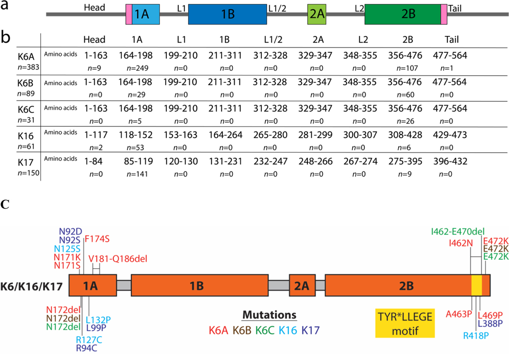
Pachyonychia congenita (PC) is a genetic disorder of keratin that presents with nail dystrophy, painful palmoplantar keratoderma, and other clinical manifestations. We investigated the genotype‒structurotype‒phenotype correlations seen with mutations in keratin genes (keratin [K]6A, K6B, K6C, K16, K17) and utilized protein structure modeling of high-frequency mutations to examine the functional importance of keratin structural domains in PC pathogenesis. Participants of the International PC Research Registry underwent genetic testing and completed a standardized survey on their symptoms. Our results support previous reports associating oral leukokeratosis with K6A mutations and cutaneous cysts, follicular hyperkeratosis, and natal teeth with K17 mutations. Painful keratoderma was prominent with K6A and K16 mutations. Nail involvement was most common in patients with K6A mutation and least common in those with K6C mutation. Across keratin subtypes, patients with coil 2B mutations had the greatest impairment in ambulation, and patients with coil 1A mutations reported more emotional issues. Molecular modeling demonstrated that hotspot missense mutations in PC largely disrupted hydrophobic interactions or surface charge. The former may destabilize keratin dimers/tetramers, whereas the latter likely interferes with higher-order keratin filament formation. Understanding the pathologic alterations in keratin structure improves our knowledge of how PC genotype correlates with clinical phenotype, advancing insight into disease pathogenesis and therapeutic development.


Conflict of interest statement

CONFLICT OF INTEREST

The authors have no conflicts of interest to disclose.

Figures

Figure 1.
Figure 1.. Common clinical manifestations of PC
include (A) toenail dystrophy, (B) plantar keratoderma, (C) cysts, and (D) oral leukokeratosis. Hypertrophic nail dystrophy is the predominant clinical feature of PC, typically presenting either with nails that grow to full length but have an upward slant caused by distal hyperkeratosis, or nails with a nail plate that terminates prematurely leaving a sloping distal region of hyperkeratosis and exposed distal finger tip. Palmoplantar keratoderma, which may be associated with intense pain, is another manifestation that typically presents when a child begins walking. Dermatological manifestations of PC include pilosebaceous cysts and follicular hyperkeratosis. Oral leukokeratosis presents as thickened white patches on the tongue and cheek. Other common manifestations of PC include natal or prenatal teeth, as well as hyperhidrosis (not pictured).
Figure 2.
Figure 2.. Mutations by KRT Gene Location
(A) Schematic of the domain structure of the keratin protein. Sequence domains were obtained from the Human Intermediate Filament Database (interfil.org). (B) The number of amino acid residues and patients with mutations located in each subdomain. (C) Mapping of PC missense mutations onto the keratin domain structure.
Figure 3.
Figure 3.. Nail involvement by KRT gene groups
(A) Fingernail involvement by KRT gene groups. Ninety-six percent of K6A patients had definite or likely fingernail involvement, while as only 3% of K6C patients did. Though most patients did not have fingernails surgically removed, K6A patients had the highest percentage (10%) of patients who had at least 1 fingernail removed. (B) Toenail involvement by KRT gene groups. Ninety-seven percent of K6A patients had definite or likely toenail involvement compared to 19% of K6C patients. K6B patients had the highest percentage (16%) of patients who had at least 1 toenail removed. *Definite involvement defined as >=7 nails reported to be thickened, likely as 4–6 nails, possible as 1–3 and none as 0.
Figure 4.
Figure 4.. Functional impairment, severity of pain, and emotional impact associated with plantar keratoderma in PC by KRT gene group and subdomain
(A) Percentage* of PC patients reporting walking with pain or inability to walk without a walking aid grouped based on location of mutations within KRT gene. (B) Absolute number and percentage* of patients reporting each level of pain by KRT gene. (C) Percentage* of PC patients reporting an emotional or psychiatric issue by KRT gene and subdomain location of mutation. *Percentages were calculated by dividing by n patients who responded for each question.
Figure 5.
Figure 5.. Molecular modeling of selected PC mutations.
Ribbon diagrams show overall structure of the K6A/K16–1A (A), K6A/K17–1A (E), and K6A/K16–2B (G) models with zoomed in view of regions containing residues involved in PC mutations (denoted by an asterisk). Comparisons of the hydrophobic surfaces of wild-type and mutated models harboring the F174SK6A (B), L99PK17 (F) and L469PK6A (I) show loss of hydrophobic surface area due to mutation. Comparisons of the charge surfaces of wild-type and mutated models harboring the N171KK6A (C), R127CK16 (D) and E472KK6A (H) mutations show significant differences in surface charge.

References

    1. Ahn J, Jo I, Kang SM, Hong S, Kim S, Jeong S, et al. Structural basis for lamin assembly at the molecular level. Nat Commun 2019;10(1):3757. - PMC - PubMed
    1. Baker NA, Sept D, Joseph S, Holst MJ, McCammon JA. Electrostatics of nanosystems: application to microtubules and the ribosome. Proc Natl Acad Sci USA. 2001;98:10037–41. - PMC - PubMed
    1. Bunick CG, Milstone LM. The x-ray crystal structure of the keratin 1-keraton 10 helix 2B heterodimer reveals molecular surface properties and biochemical insights into human skin disease. J Invest Dermatol. 2017;137(1):142–150. - PMC - PubMed
    1. Chamcheu JC, Siddiqui IA, Syed DN, Adhami VM, Liovic M, Mukhtar H. Keratin Gene Mutations in Disorders of Human Skin and its Appendages. Arch Biochem Biophys. 2011;508:123–137. - PMC - PubMed
    1. Connors JB, Rahil AK, Smith FJ, McLean WH, Milstone LM. Delayed-onset pachyonychia congenita associated with a novel mutation in the central 2B domain of keratin 16. Br J Dermatol. 2001;144:1058–62. - PubMed

Publication types