Review
. 2021 Sep:105:103144.
doi: 10.1016/j.dnarep.2021.103144. Epub 2021 Jun 3.

ADP-ribosylation of DNA and RNA

Affiliations
Review

ADP-ribosylation of DNA and RNA

Joséphine Groslambert et al. DNA Repair (Amst). 2021 Sep.

Abstract

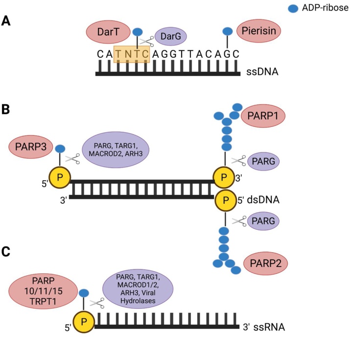
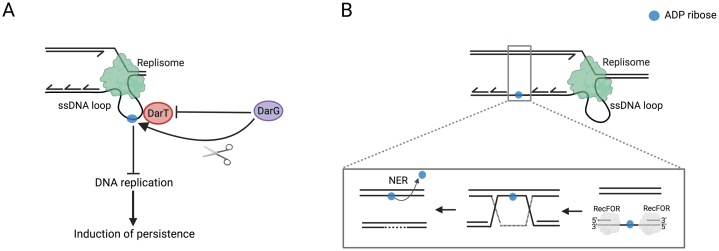
ADP-ribosylation is a chemical modification of macromolecules found across all domains of life and known to regulate a variety of cellular processes. Notably, it has a well-established role in the DNA damage response. While it was historically known as a post-translational modification of proteins, recent studies have shown that nucleic acids can also serve as substrates of reversible ADP-ribosylation. More precisely, ADP-ribosylation of DNA bases, phosphorylated DNA ends and phosphorylated RNA ends have been reported. We will discuss these three types of modification in details. In a variety of bacterial species, including Mycobacterium tuberculosis, ADP-ribosylation of thymidine has emerged as the mode of action of a toxin-antitoxin system named DarTG, with the resultant products perceived as DNA damage by the cell. On the other hand, mammalian DNA damage sensors PARP1, PARP2 and PARP3 were shown to ADP-ribosylate phosphorylated ends of double-stranded DNA in vitro. Additionally, TRPT1 and several PARP enzymes, including PARP10, can add ADP-ribose to the 5'-phosphorylated end of single-stranded RNA in vitro, representing a novel RNA capping mechanism. Together, these discoveries have led to the emergence of a new and exciting research area, namely DNA and RNA ADP-ribosylation, that is likely to have far-reaching implications for the fields of DNA repair, replication and epigenetics.

Keywords: ADP-ribosylation; DNA damage response; DNA modification; PARP; RNA modification.


Conflict of interest statement

The authors report no declarations of interest.

Figures

Fig. 1
Fig. 1
ADP-ribosylation of nucleic acids. (A) ADP-ribosylation of DNA bases. DarT MARylates the second thymine base of a TNTC motif on ssDNA and this modification can be removed by DarG. Pierisin from the cabbage butterfly and its orthologue MARylate guanine on ssDNA in an irreversible manner. (B) ADP-ribosylation of DNA ends. PARP1 and PARP2 PARylate phosphorylated termini on dsDNA with a preference for the 3’- and 5’-terminal phosphate, respectively. The PARP1/PARP2-mediated DNA modification can be removed by PARG. PARP3 MARylates phosphorylated termini on dsDNA with a preference for the 5’-terminal phosphate. This modification can be reversed by PARG, TARG1, MacroD2 and ARH3. (C) ADP-ribosylation of RNA ends. PARP10/PARP11/PARP15 and TRPT1 MARylate ssRNA at the 5’-terminus, forming a non-canonical cap. PARP10 was also shown to modify the 3’-terminal phosphate of ssRNA, albeit less efficiently than at the 5’-terminus. The modification catalysed by PARP10 and TRPT1 was shown to be reversed by PARG, TARG1, MacroD1, MacroD2 and ARH3. Viral macrodomain-containing hydrolases could reverse the modification catalysed by PARP10 on ssRNA 5’-phosphorylated ends.
Fig. 2
Fig. 2
Repair of DarT-catalysed DNA ADP-ribosylation. (A) Direct reversal of DarT-induced DNA lesion by DarG. DarG also inhibits the catalytic activity of DarT through binding and physically sequestering the enzyme. The DarT-mediated DNA lesion inhibits replication which could induce persistence. (B) Model for the DarG-independent repair of DarT-induced DNA lesion. DarT ADP-ribosylates ssDNA loops arising during replication, leading to the generation of single-stranded gaps (SSG). The RecF-mediated homologous recombination repair pathway recognizes the SSG and repairs the gap. The ADP-ribosylated DNA adduct (blue dot) is then repaired by the nucleotide excision repair (NER) pathway.

References

    1. Hoeijmakers J.H.J. DNA damage, aging, and Cancer. N. Engl. J. Med. 2009 doi: 10.1056/nejmra0804615. - DOI - PubMed
    1. Perina D., Mikoč A., Ahel J., Ćetković H., Žaja R., Ahel I. Distribution of protein poly(ADP-ribosyl)ation systems across all domains of life. DNA Repair (Amst). 2014 doi: 10.1016/j.dnarep.2014.05.003. - DOI - PMC - PubMed
    1. Gibson B.A., Kraus W.L. New insights into the molecular and cellular functions of poly(ADP-ribose) and PARPs. Nat. Rev. Mol. Cell Biol. 2012 doi: 10.1038/nrm3376. - DOI - PubMed
    1. Martin-Hernandez K., Rodriguez-Vargas J.M., Schreiber V., Dantzer F. Expanding functions of ADP-ribosylation in the maintenance of genome integrity. Semin. Cell Dev. Biol. 2017 doi: 10.1016/j.semcdb.2016.09.009. - DOI - PubMed
    1. Lüscher B., Bütepage M., Eckei L., Krieg S., Verheugd P., Shilton B.H. ADP-Ribosylation, a Multifaceted Posttranslational Modification Involved in the Control of Cell Physiology in Health and Disease. Chem. Rev. 2018 doi: 10.1021/acs.chemrev.7b00122. - DOI - PubMed

Publication types

LinkOut - more resources