Case Reports
. 2021 Jun 11;14(6):e242855.
doi: 10.1136/bcr-2021-242855.

Pneumocephalus with meningitis secondary to an old traumatic anterior cranial fossa defect

Affiliations
Case Reports

Pneumocephalus with meningitis secondary to an old traumatic anterior cranial fossa defect

Freston Marc Sirur et al. BMJ Case Rep. .

Abstract

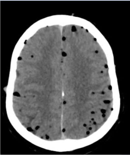
We report a case of a 30-year-old man who presented with altered mental status, fever, headache and vomiting for 3 days. An initial CT scan of the brain revealed the presence of pneumocephalus with a bony defect in the anterior cranial fossa. The pneumocephalus was not explained initially and the patient was re-examined for any signs of trauma to the face, and a review of the history revealed a series of three traumatic events months prior to this illness. Further laboratory studies revealed Streptococcus pneumoniae in the blood and bacterial meningitis. He was treated with antibiotics and was later taken up for endoscopic repair of the skull base defect. This case highlights the importance of recognising post-traumatic pneumocephalus with superimposed meningitis and sepsis months after a traumatic event to the skull base.

Keywords: emergency medicine; infection (neurology); meningitis; otolaryngology / ENT; trauma.


Conflict of interest statement

Competing interests: None declared.

Figures

Figure 1
Figure 1
Sequence of events-presentation, diagnostics and treatment. HR - heart rate; BP - blood pressure; GCS - Glasgow Coma Scale; ABCDE - Airway, Breathing, Circulation, Disability, Exposure; 3D - three dimensional; HRCT - High resolution CT; ICU - intensive care unit; URTI - upper respiratory tract infection;CSF-cerebrospinal fluid; IV- intravenous3
Figure 2
Figure 2
Initial CT of the brain (axial section) showing ‘air bubble’ sign.
Figure 3
Figure 3
CT of the face with three-dimensional reconfiguration showing a defect measuring ~3.8 mm in the floor of the anterior cranial fossa along left with focal herniation of the brain parenchyma through the defect.
Figure 4
Figure 4
Axial T2-FLAIR (Fluid attenuated inversion recovery) and diffusion-weighted MR images show subcortical white matter patchy hyper-intensities on FLAIR sequences and showing restricted diffusion on DWI (diffusion weighted imaging) sequences in bilateral frontal lobes.

References

    1. Pillai P, Sharma R, MacKenzie L, et al. . Traumatic tension pneumocephalus - Two cases and comprehensive review of literature. Int J Crit Illn Inj Sci 2017;7:58. 10.4103/IJCIIS.IJCIIS_8_17 - DOI - PMC - PubMed
    1. Markham JW. The clinical features of pneumocephalus based upon a survey of 284 cases with report of 11 additional cases. Acta Neurochir 1967;16:1–78. 10.1007/BF01401900 - DOI - PubMed
    1. Orebaugh SL, Margolis JH. Post-Traumatic intracerebral pneumatocele: case report. J Trauma 1990;30:1577–80. 10.1097/00005373-199012000-00025 - DOI - PubMed
    1. Mendelsohn DB, Hertzanu Y. Intracerebral pneumatoceles following facial trauma: CT findings. Radiology 1985;154:115–8. 10.1148/radiology.154.1.3964928 - DOI - PubMed
    1. Cunqueiro A, Scheinfeld MH. Causes of pneumocephalus and when to be concerned about it. Emerg Radiol 2018;25:331–40. 10.1007/s10140-018-1595-x - DOI - PubMed

Publication types