Review
. 2021 Jul 1;35(13-14):940-962.
doi: 10.1101/gad.348523.121. Epub 2021 Jun 11.

The biological underpinnings of therapeutic resistance in pancreatic cancer

Affiliations
Review

The biological underpinnings of therapeutic resistance in pancreatic cancer

Gregory L Beatty et al. Genes Dev. .

Abstract

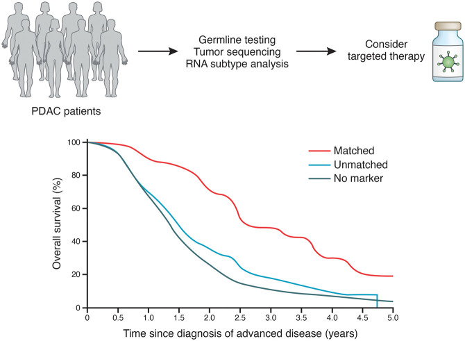
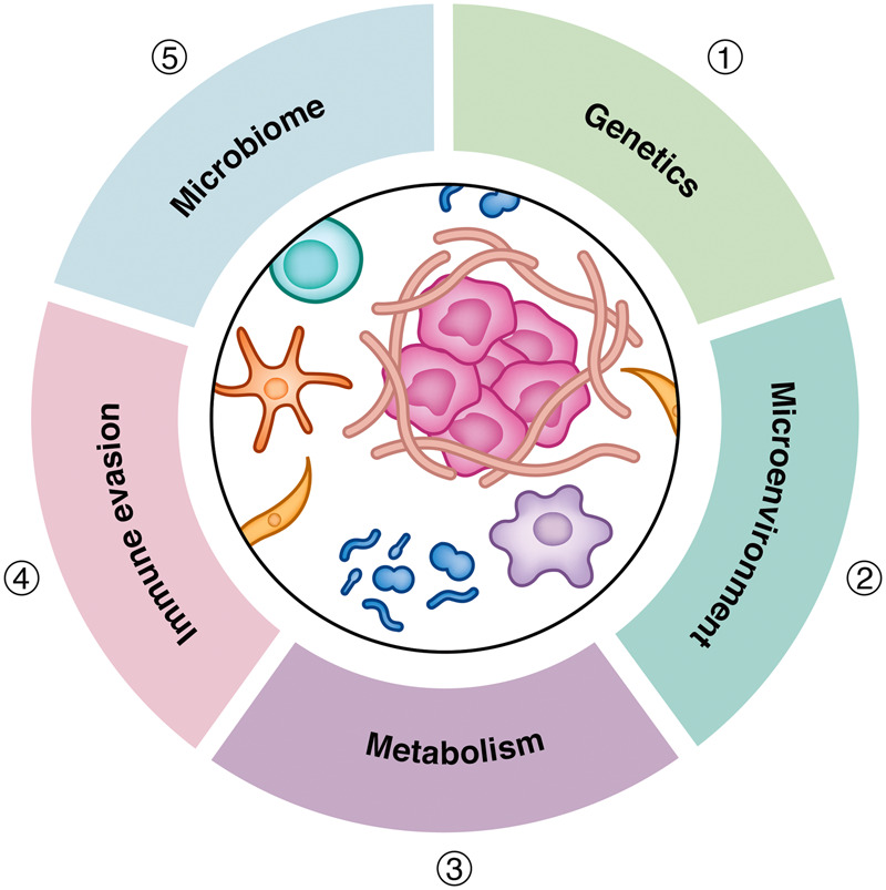
Pancreatic ductal adenocarcinoma (PDAC) is a leading cause of cancer-related mortality in the United States and has only recently achieved a 5-yr survival rate of 10%. This dismal prognosis reflects the remarkable capacity of PDAC to effectively adapt to and resist therapeutic intervention. In this review, we discuss recent advances in our understanding of the biological underpinnings of PDAC and their implications as targetable vulnerabilities in this highly lethal disease.

Keywords: PDAC; genetics; metabolism; microbiome; pancreatic cancer; pancreatic tumor microenvironment; targeted therapy; therapeutic resistance.


Figures

Figure 1.
Figure 1.
Precision medicine in pancreatic cancer. (Top) PDAC patients exhibit a breadth of tumor biology with generally poor responses to standard chemotherapy. Precision medicine, rooted in a multiomic approach to tumor/patient sequencing, can uncover potential avenues for targeted therapies. (Bottom) Adoption of a precision medicine approach coupled with matched therapies can substantially improve survival of patients with pancreatic cancer. (Bottom figure adapted from Pishvaian et al. 2020, © 2020, with permission from Elsevier.)
Figure 2.
Figure 2.
Metabolic mechanisms of therapeutic resistance in PDA. (A) Cell-autonomous mechanisms of therapeutic resistance. PDAC cells enhance the production of nucleic acids from glucose through the pentose phosphate pathway (PPP) to promote resistance to gemcitabine. Macropinocytosis and autophagy provide nutrients (e.g. Fe2+, amino acids) to support biosynthesis and survival. Autophagy also removes MHC-I from the cell surface to impair recognition by the antitumor immune system. Malic enzyme 1 (ME1)-derived NADPH and the nuclear factor erythroid 2-related factor 2 (NRF2) pathway promote resistance to reactive oxygen species (ROS). NRF2 is transcriptionally activated by mutant KRAS and post-translationally stabilized by the ataxia-telangiectasia group D-associated protein (ATDC)-mediated binding and inhibition of Kelch-like ECH-associated protein 1 (KEAP1). (B) Tumor microenvironment-mediated mechanisms of therapeutic resistance. Deoxycytidine (dC) derived from cancer-associated fibroblasts (CAFs) and tumor-associated macrophages (TAMs) promotes resistance to gemcitabine. Pyruvate derived from CAFs and circulating asparagine (Asn) promote resistance to mitochondrial inhibitors.
Figure 3.
Figure 3.
Stromal determinants and therapeutic challenges to intervening on PDAC oathogenesis. The stromal compartment in PDAC is shaped by tumor-infiltrating leukocytes recruited from the peripheral blood. These leukocytes coordinate therapeutic resistance and aid in PDAC progression and metastasis. Shown are therapeutic challenges established by this dynamic interaction between host and tumor. (1) Within the peripheral blood, deficiencies in immune health are observed that may limit the efficacy of immunotherapy. (2) Tumors recruit immune-suppressive cells and exclude effector T cells. (3) Within tumors, genetic aberrations in the cancer cells instruct the formation of a microenvironment marked by immunosuppression, nutrient deprivation, and a desmoplastic reaction. (4) The tumor microenvironment supports the metastatic cascade. (5) Tumors produce factors that alter host physiology and condition distant organs for increased metastatic susceptibility.
Figure 4.
Figure 4.
Overview over the pleotropic effects of the microbiome on the pancreatic tumor microenvironment. (1) TLR ligation by pancreas-intrinsic bacteria-derived peptides skews the immune system toward a tolerogenic phenotype. (2) Bacterial metabolites and peptides from the gut microbiome further promote a tolerogenic immune infiltrate. (3) Bacteria produce cytidine deaminase, which metabolizes gemcitabine to its inactive form difluorodeoxyuridine (dFdU). (4) Digestive juices and antimicrobioal peptides (AMPs) shape the intestinal flora, which then influence the pancreatic TME. (5) Bacterial peptides with similarity to human proteins may trigger cognate T-cell priming and activation of antitumor immunity.
Figure 5.
Figure 5.
Key determinants of therapeutic resistance in pancreatic cancer. Therapeutic resistance in pancreatic ductal adenocarcinoma is influenced by a myriad of biological pathways directed by (1) genetics, including alterations in oncogenes and tumor suppressor genes; (2) the microenvironment, including fibrosis and poor vascularity, which are hallmarks of pancreatic cancer and contribute to limit drug delivery and impact the contexture of the host immune response; (3) metabolism, including metabolites that shape tumor and host biology as well as cancer cell sensitivity to cytotoxic agents; (4) immune evasion, the capacity to avoid detection and elimination by T cells and other effector immune cell populations; and (5) the microbiome, including gut and intratumoral microbes as well as their byproducts.

Similar articles

Cited by

References

    1. Adams CR, Htwe HH, Marsh T, Wang AL, Montoya ML, Subbaraj L, Tward AD, Bardeesy N, Perera RM. 2019. Transcriptional control of subtype switching ensures adaptation and growth of pancreatic cancer. Elife 8: e45313. 10.7554/eLife.45313 - DOI - PMC - PubMed
    1. Aguirre AJ, Nowak JA, Camarda ND, Moffitt RA, Ghazani AA, Hazar-Rethinam M, Raghavan S, Kim J, Brais LK, Ragon D, et al. 2018. Real-time genomic characterization of advanced pancreatic cancer to enable precision medicine. Cancer Discov 8: 1096–1111. 10.1158/2159-8290.CD-18-0275 - DOI - PMC - PubMed
    1. Alcalá S, Sancho P, Martinelli P, Navarro D, Pedrero C, Martín-Hijano L, Valle S, Earl J, Rodríguez-Serrano M, Ruiz-Cañas L, et al. 2020. ISG15 and ISGylation is required for pancreatic cancer stem cell mitophagy and metabolic plasticity. Nat Commun 11: 2682. 10.1038/s41467-020-16395-2 - DOI - PMC - PubMed
    1. Alistar A, Morris BB, Desnoyer R, Klepin HD, Hosseinzadeh K, Clark C, Cameron A, Leyendecker J, D'Agostino R, Topaloglu U, et al. 2017. Safety and tolerability of the first-in-class agent CPI-613 in combination with modified FOLFIRINOX in patients with metastatic pancreatic cancer: a single-centre, open-label, dose-escalation, phase 1 trial. Lancet Oncol 18: 770–778. 10.1016/S1470-2045(17)30314-5 - DOI - PMC - PubMed
    1. Ashton TM, McKenna WG, Kunz-Schughart LA, Higgins GS. 2018. Oxidative phosphorylation as an emerging target in cancer therapy. Clin Cancer Res 24: 2482–2490. 10.1158/1078-0432.CCR-17-3070 - DOI - PubMed

Publication types

MeSH terms