. 2021 Jun;9(6):e002508.
doi: 10.1136/jitc-2021-002508.

Mesoporous silica nanoparticles inflame tumors to overcome anti-PD-1 resistance through TLR4-NFκB axis

Affiliations

Mesoporous silica nanoparticles inflame tumors to overcome anti-PD-1 resistance through TLR4-NFκB axis

Mayu Sun et al. J Immunother Cancer. 2021 Jun.

Abstract

Background: The clinical benefits of antiprogrammed cell death protein 1 (PD-1) therapy are compromised by resistance in immunologically cold tumors. Convergence of immunotherapy and bioengineering is potential to overcome the resistance. Mesoporous silica nanoparticles (MSNs) are considered the most promising inorganic biological nanomaterials for clinical transformation, however, the fundamental influence of MSNs on immunotherapy is unclear. In this study, we aimed to investigate the role of MSNs in tumor resensitization and explore the feasibility of MSNs combined with anti-PD-1 in cancer therapy.

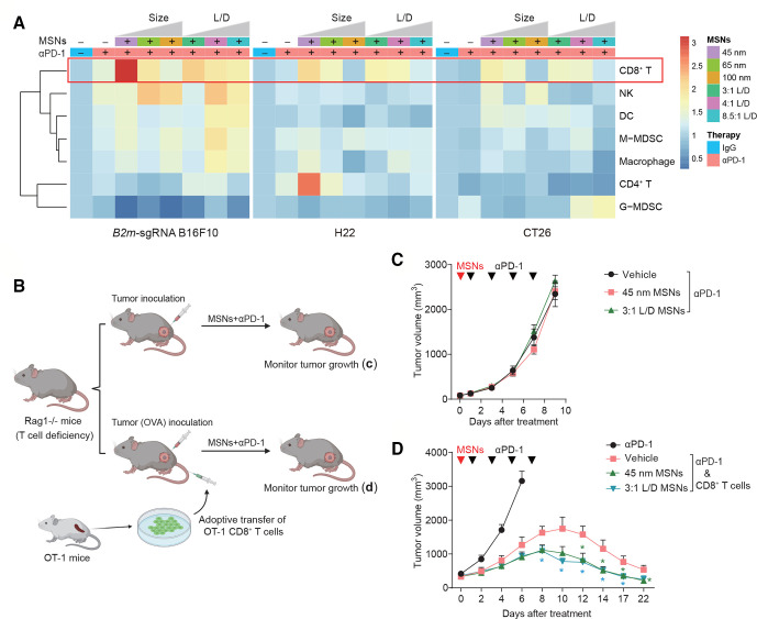
Methods: Intrinsic and acquired resistant tumors, as well as spontaneous and secondary tumor recurrence models, were used to evaluate the influence of MSNs and the synergistical effect with anti-PD-1 therapy. The roles of CD8+ cytotoxic T-lymphocytes (CTLs) and macrophages were assessed in Rag-1-/- mice, ovalbumin/OT-1 TCR transgenic T-cell system, and other blocking mice models. Mechanistic studies were processed by transcriptomics analysis and conducted in primary cells, in vitro coculture systems, and Toll-like receptor 4 (TLR4) knockout mice.

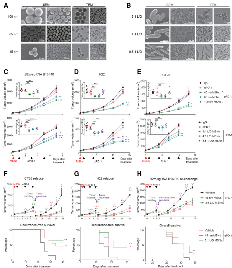
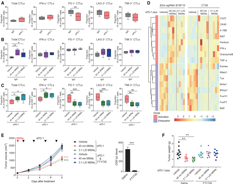
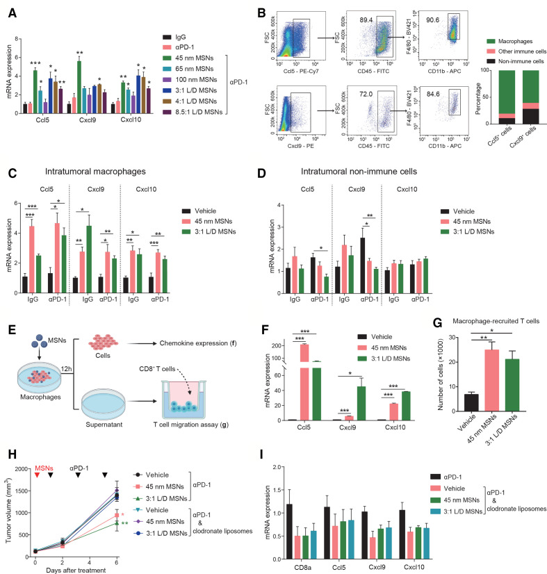
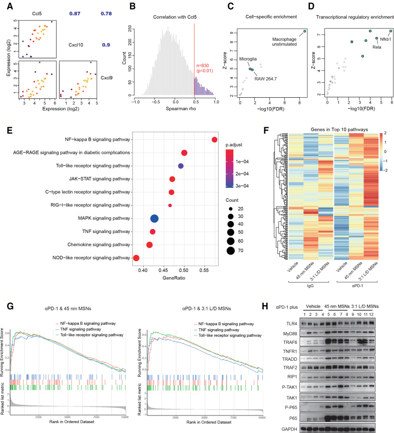
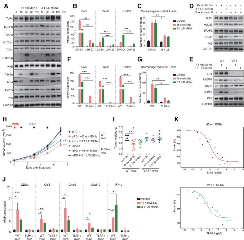
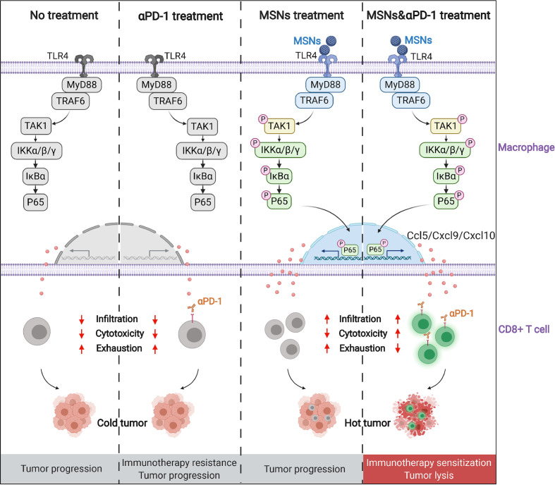
Results: Both granular and rod-shaped MSNs efficiently overcame tumor resistance with dependence on diameter and aspect ratio. Only once injection of MSNs in prior to anti-PD-1 markedly improved the treatment efficacy, protective immunity, and prognosis. MSNs per se boosted infiltration of CTLs as the early event (days 2-3); and synergistically with anti-PD-1 therapy, MSNs rapidly established a T cell-inflamed microenvironment with abundant high-activated (interferon-γ/tumor necrosis factor-α/Perforin/GranzymeB) and low-exhausted (PD-1/lymphocyte-activation gene 3 (LAG-3)/T-cell immunoglobulin and mucin-domain containing-3 (TIM-3)) CTLs. Chemokines Ccl5/Cxcl9/Cxcl10, which were produced predominantly by macrophages, promoted MSNs-induced CTLs infiltration. MSNs led to high Ccl5/Cxcl9/Cxcl10 production in vitro and in mice through regulating TLR4-NFκB axis. Blocking TLR4-NFκB axis in macrophages or CTLs infiltration abrogated MSNs-induced resensitization to anti-PD-1 therapy.

Conclusions: MSNs efficiently and rapidly inflame immunologically cold tumors and resensitize them to anti-PD-1 therapy through TLR4-NFκB-Ccl5/Cxcl9/Cxcl10 axis. MSNs-based theranostic agents can serve as sensitizers for patients with resistant tumors to improve immunotherapy.

Keywords: combined modality therapy; immunotherapy; lymphocytes; macrophages; programmed cell death 1 receptor; tumor-infiltrating.


Conflict of interest statement

Competing interests: None declared.

Figures

Figure 1
Figure 1
MSNs overcome resistance and prevent relapse of αPD-1-resistant tumors. (A) SEM and TEM images of spherical MSNs with diameters of 100, 65 and 45 nm. (B) SEM and TEM images of rod-like MSNs with aspect ratios of 3:1, 4:1, 8.5:1. (C–E) Tumor growth and tumor weights of B2m-sgRNA B16F10 (C), H22 (D) and CT26 (E) tumor-bearing mice treated with different MSNs treatment (top: sizes; bottom: L/Ds) prior to αPD-1. (F, G) Growth of recurrent tumors (top) and recurrence-free survival (bottom) of tumor-bearing mice after indicated treatments. (H) Growth of rechallenged tumors (top) and overall survival (bottom) of B2m-sgRNA B16F10 tumor-bearing mice after indicated treatments. Error bars represent mean±SEM (n>6 per group in C–H). P value for tumor measurement was calculated by unpaired Student’s t-test. P value for survival curve was calculated by the log-rank (Mantel-Cox) test. (*P<0.05, **p<0.01, ***p<0.001). MSNs, mesoporous silica nanoparticles; PD-1, programmed cell death protein 1; TEM, tumor microenvironment.
Figure 2
Figure 2
MSNs resensitize tumors to αPD-1 through CTLs. (A) Heatmap showing the proportion of tumor-infiltrating CD8+ T cells, NKs, DCs, M-MDSCs, macrophages, CD4+ T cells and G-MDSCs in the Tme after indicated treatments. (B) Scheme of tumor therapy in T cell-deficiency Rag1-/- mice and T cell-regained mice by transferring. (C) Growth of B2m-sgRNA B16F10 tumors in Rag1-/- mice after indicated treatments. (D) Growth of B16F10-OVA tumors in Rag1-/- mice with adoptive transfer of OT-1 CTLs. Error bars represent mean±SEM (n>5 per group in (C, D)). P value was calculated by unpaired Student’s t-test (*P<0.05, **p<0.01, ***p<0.001). CTLs, cytotoxic T-lymphocytes; DCs, dendritic cells; G-MDSCs, granulocytic myeloid-derived suppressor cells; M-MDSCs, monocytic MDSCs; MSNs, mesoporous silica nanoparticles; NKs, natural killer; OVA, ovalbumin; PD-1, programmed cell death protein 1.
Figure 3
Figure 3
MSNs drive CTLs infiltration to promote CTLs activation synergistically with αPD-1 at the beginning of treatment. (A–C) On day 2–3 after indicated treatments, the proportion of total CTLs and INF-γ, PD-1, LAG-3 and Tim-3 positive CTLs in B2m-sgRNA B16F10 tumor tissues was quantified by flow cytometry. (D) On day 3 after indicated treatments, the mRNA expression in CTLs isolated from B2m-sgRNA B16F10 or CT26 tumor tissues was shown. (E, F) Tumor growth (E) and tumor weights (F) of B2m-sgRNA B16F10 tumor-bearing mice treated with FTY720 to block T cell infiltration. Control mice received saline. Flow cytometry analysis quantified the numbers of CTLs collected from blood 3 days postinitial FTY720 administration. Error bars represent mean±SEM (n>6 per group in E). P value was calculated by unpaired Student’s t-test (*p<0.05, **p<0.01, ***p<0.001). CTLs, cytotoxic T-lymphocytes; INF-γ, interferon-γ; MSNs, mesoporous silica nanoparticles; PD-1, programmed cell death protein 1; TNFα, tumor necrosis factor-α.
Figure 4
Figure 4
MSNs promote the production of T cell-recruitment chemokines in macrophages. (A) The mRNA expression of CCL5, CXCL9, and CXCL10 in B2m-sgRNA B16F10 tumor tissues after indicated treatments was determined by qRT-PCR. (B) The major cell types that produced CCL5 and CXCL9 in B2m-sgRNA B16F10 tumor tissues were analyzed by flow cytometry. the right graph quantified the percentage of CCL5 and CXCL9 producing cell types. (C, D) The mRNA expression of CCL5, CXCL9 and CXCL10 in intratumoral macrophages (CD11b+F4/80+) (C) and non-immune cells (CD45) (D) isolated from B2m-sgRNA B16F10 tumor tissues was determined by qRT-PCR. (E) Scheme of in vitro macrophages treatment and coincubation. RAW 264.7 cells were treated by 100 µg/mL MSNs for 12 hours. Cells were collected for gene expression assay, and supernatant was collected for T cell recruitment assay. (F) The mRNA expression of CCL5, CXCL9 and CXCL10 in RAW 264.7 cells after MSNs treatments was determined by qRT-PCR. (G) Quantification of migrated T cells recruited by the supernatant of macrophages after MSNs treatments. (H) Tumor growth of B2m-sgRNA B16F10 tumor-bearing mice treated with clodronate liposomes to deplete macrophages. Control mice received dimethyl sulfoxide. (I) The mRNA expression of CD8a, CCL5, CXCL9 and CXCL10 in tumor tissues after indicated treatments was determined by qRT-PCR. Error bars represent mean±SEM (n>7 per group in H). P value was calculated by unpaired Student’s t-test. (*P<0.05, **p<0.01, ***p<0.001). MSNs, mesoporous silica nanoparticles; PD-1, programmed cell death protein 1; FSC, forward scatter.
Figure 5
Figure 5
T cell-recruitment chemokines expression is associated with NF-κB signaling after MSNs treatment. (A) Correlation among the expression of T cell-recruitment chemokines across all B2m-sgRNA B16F10 tumor tissues. (B) Through transcriptomic analysis, all genes with significantly positive correlation with CCL5 were identified. (C) Cell-specific enrichment of CCL5-correlated genes. (D) Transcriptional regulatory enrichment of CCL5-correlated genes. (E) Top 10 signaling pathways by KEGG pathway enrichment of Ccl5-correlated genes. (F) In MSNs-treated tumors, the expression of genes in the top 10 Ccl5-related signaling pathways was presented by heatmap. (G) In tumor tissues, the activation changes of NF-κB, TNF and TLR signaling pathways in αPD-1 plus MSNs groups compared with αPD-1 alone were revealed by GSEA. (H) In tumor tissues after indicated treatments, the levels of proteins in TLR4, TNFR and NF-κB signaling were determined by Western blot. GSEA, Gene Set Enrichment Analysis; MSNs, mesoporous silica nanoparticles; PD-1, programmed cell death protein 1; TLR, Toll-like receptor; TNF, tumor necrosis factor.
Figure 6
Figure 6
MSNs overcome tumor resistance through activating TLR4/NF-κB signaling in macrophages. (A) RAW 264.7 cells were treated with 100 µg/mL MSNs for different times and the protein levels were determined by Western blot. (B) RAW 264.7 cells were treated with 10 µM JSH-23 for 3 hours and then 100 µg/mL MSNs for another 12 hours. the mRNA expression of CCL5, CXCL9 and CXCL10 was determined by qRT-PCR. (C) RAW 264.7 cells were treated with 10 µM JSH-23 for 3 hours and then 100 µg/mL MSNs for another 12 hours. T cells recruited by the supernatant of macrophages were quantified through migration assay. (D) RAW 264.7 cells were treated with 100 µM TLR4 blockade sparstolonin B for 3 hours and then 100 µg/mL MSNs treatment for 1 hour. The protein levels were determined by Western blot. (E) Bone marrow-derived macrophages (BMDMs) from wide type (WT) and TLR4-deficient (TLR4-/-) mice were treated with 100 µg/mL MSNs for 1 hour. The protein levels were determined by Western blot. (F) WT and TLR4-/- BMDMs were treated 100 µg/mL MSNs treatment for 12 hours. The mRNA expression of CCL5, CXCL9 and CXCL10 was determined by qRT-PCR. (G) WT and TLR4-/- BMDMs were treated 100 µg/mL MSNs treatment for 12 hours. T cells recruited by the supernatant of macrophages were quantified through migration assay. (H, I) Tumor growth (H) and tumor weights (I) of B2m-sgRNA B16F10 tumors in WT and TLR4-/- mice after indicated treatments. (J) After indicated treatments, the mRNA expression of CD8a, CCL5, CXCL9, CXCL10 and IFN-γ in tumor tissues was determined by qRT-PCR. Error bars represent mean±SEM (n>5 per group in H). (K) The dose-response curves of MSNs with TLR4 in MST affinity analysis. The concentrations were 0.2 mg/mL for FITC-labeled MSNs and 0.1 nM–2.5 μM for TLR4 protein. P value was calculated by unpaired Student’s t-test. (*P<0.05, **p<0.01, ***p<0.001). MSNs, mesoporous silica nanoparticles; IFN-γ, interferon-γ; TLR4, Toll-like receptor 4.
Figure 7
Figure 7
Schematic depiction of the synergistical effect of MSNs and PD-1 blockade on tumor immunotherapy. MSNs, mesoporous silica nanoparticles; PD-1, programmed cell death protein 1; TLR4, Toll-like receptor 4.

References

    1. Sharpe AH, Wherry EJ, Ahmed R, et al. . The function of programmed cell death 1 and its ligands in regulating autoimmunity and infection. Nat Immunol 2007;8:239–45. 10.1038/ni1443 - DOI - PubMed
    1. Wherry EJ, Kurachi M. Molecular and cellular insights into T cell exhaustion. Nat Rev Immunol 2015;15:486–99. 10.1038/nri3862 - DOI - PMC - PubMed
    1. Sharma P, Hu-Lieskovan S, Wargo JA, et al. . Primary, adaptive, and acquired resistance to cancer immunotherapy. Cell 2017;168:707–23. 10.1016/j.cell.2017.01.017 - DOI - PMC - PubMed
    1. Reck M, Rodríguez-Abreu D, Robinson AG, et al. . Pembrolizumab versus chemotherapy for PD-L1-positive non-small-cell lung cancer. N Engl J Med 2016;375:1823–33. 10.1056/NEJMoa1606774 - DOI - PubMed
    1. Bonaventura P, Shekarian T, Alcazer V, et al. . Cold tumors: a therapeutic challenge for immunotherapy. Front Immunol 2019;10:168. 10.3389/fimmu.2019.00168 - DOI - PMC - PubMed

Publication types