. 2021 Jun 11;4(1):724.
doi: 10.1038/s42003-021-02250-7.

Structural basis of coronavirus E protein interactions with human PALS1 PDZ domain

Affiliations

Structural basis of coronavirus E protein interactions with human PALS1 PDZ domain

Airah Javorsky et al. Commun Biol. .

Abstract

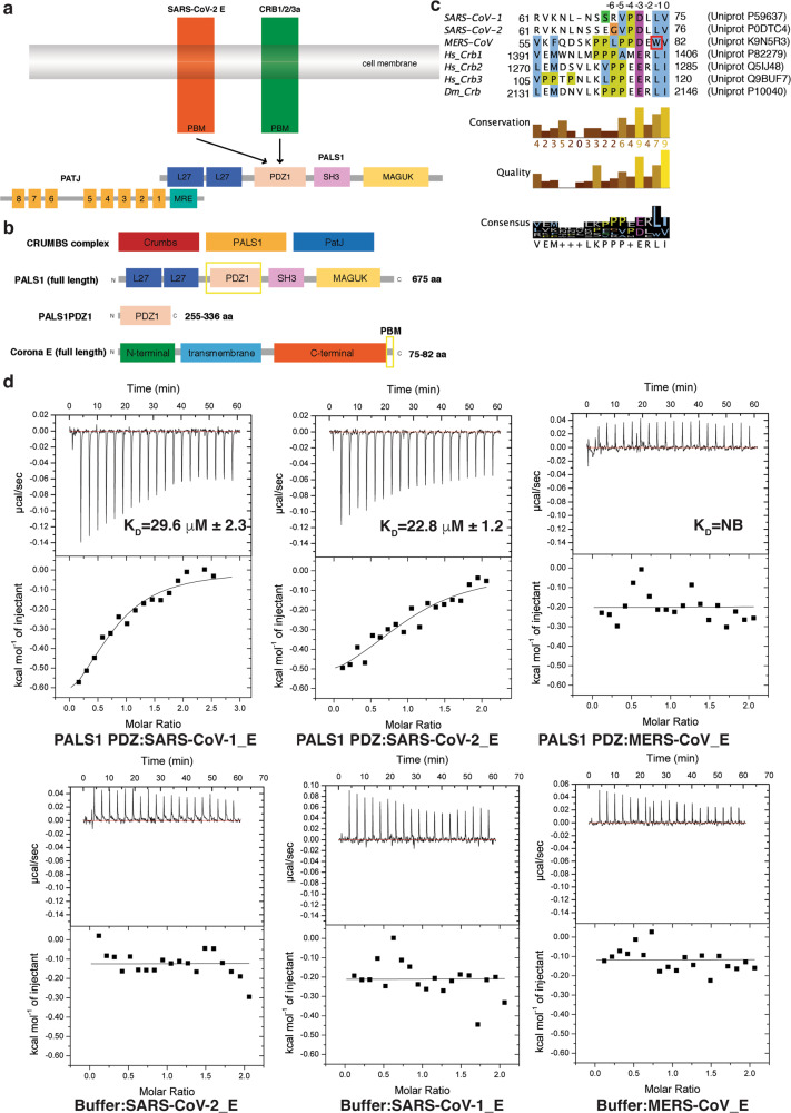
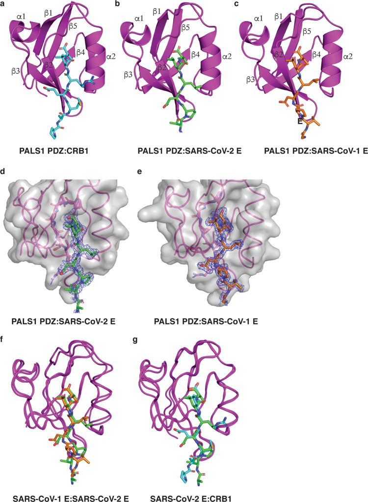
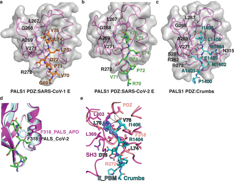
SARS-CoV-2 infection leads to coronavirus disease 2019 (COVID-19), which is associated with severe and life-threatening pneumonia and respiratory failure. However, the molecular basis of these symptoms remains unclear. SARS-CoV-1 E protein interferes with control of cell polarity and cell-cell junction integrity in human epithelial cells by binding to the PALS1 PDZ domain, a key component of the Crumbs polarity complex. We show that C-terminal PDZ binding motifs of SARS-CoV-1 and SARS-CoV-2 E proteins bind the PALS1 PDZ domain with 29.6 and 22.8 μM affinity, whereas the related sequence from MERS-CoV did not bind. We then determined crystal structures of PALS1 PDZ domain bound to both SARS-CoV-1 and SARS-CoV-2 E protein PDZ binding motifs. Our findings establish the structural basis for SARS-CoV-1/2 mediated subversion of Crumbs polarity signalling and serve as a platform for the development of small molecule inhibitors to suppress SARS-CoV-1/2 mediated disruption of polarity signalling in epithelial cells.


Conflict of interest statement

The authors declare no competing interests.

Figures

Fig. 1
Fig. 1. Interactions of PALS1 PDZ domain with SARS-CoV-1, SARS-CoV-2 and MERS-CoV E protein PBM.
a Schematic outline of Crumb’s complex interactions. b Domain organization of PALS1 and SARS-CoV-2 E protein. c Sequence alignment of C-terminal PBM motifs of coronavirus E proteins with human (hs) crumbs isoforms and D. melanogaster (dm) crumbs. Trp81 in the MERS CoV E protein is boxed in red. PBM sequences are numbered above the alignment with the first C-terminal residue designated as 0, with preceding residues numbered −1, −2, −3 etc. Uniprot accession numbers and sequence boundaries are listed for all sequences. Residue colouring is as follows: blue for hydrophobic, magenta for a negative charge, red for positive charge, yellow for prolines and white for unconserved. Three different conservation metrics calculated by Jalview are shown. Quality score is calculated for each column in an alignment by summing, for all mutations, the ratio of the two BLOSUM 62 scores for a mutation pair and each residue’s conserved BLOSUM62 score, and then plotted on a scale from 0 to 1. The consensus displayed below the alignment is the percentage of the modal residue per column. The alignment was generated using Clustal Omega followed by analysis using Jalview. d Binding profiles of isolated PALS1 PDZ domain interaction with coronavirus E protein PBM peptides are displayed. Each profile is represented by a raw thermogram (top panel) and a binding isotherm fitted with a one-site binding model (bottom panels). KD dissociation constant, ± standard deviation, NB no binding. Each of the values was calculated from at least three independent experiments. Top panels show peptide into protein titrations, bottom panels show peptide into buffer control titrations.
Fig. 2
Fig. 2. The crystal structures of PALS1 PDZ bound to SARS-CoV-1 and SARS-CoV-2 E protein PBM.
The E protein PBM peptide engages PALS1 PDZ via a shallow groove located between the β2 and α2. a Human PALS1 PDZ (magenta) is shown as a cartoon with a human CRB1 peptide (cyan) represented as sticks (PDB ID 4UU5). b PALS1 PDZ (magenta) is shown as a cartoon with SARS-CoV-2 E protein PBM (green) represented as sticks. c PALS1 PDZ (magenta) is shown as a cartoon with SARS-CoV-1 E protein PBM (orange) represented as sticks. d, e Electron density maps centred on the SARS-CoV-2 E protein PBM peptide (green) or SARS-CoV-1 protein PBM peptide (orange) in the binding groove of PALS1 PDZ. The electron density map is a 2Fo–Fc map shown as a blue mesh contoured at 1.5σ. f Overlay of ribbon traces of PALS1 PDZ with SARS-CoV-2 E protein PBM (green) and PALS1 PDZ with SARS-CoV-1 E protein PBM peptide (orange). g Overlay of ribbon traces of PALS1 PDZ with SARS-CoV-2 E protein PBM (green) and human Crumbs peptide (cyan) peptide (PDB ID 4UU5).
Fig. 3
Fig. 3. Detailed view into the interfaces of PALS 1 PDZ bound to SARS-CoV-1 and SARS-CoV-2 E protein PBM.
ac Detailed view of interactions in the binding groove of PALS1 PDZ in complex with a, b SARS-CoV-2 E protein PBM peptide (green) or SARS-CoV-1 E protein PBM peptide (orange) or c Crumbs PBM peptide (cyan) (PDB ID 4UU5). Residues involved in hydrogen bonds or ionic interactions (shown as black dotted lines) are labelled. d Superimposition of PALS1 PDZ:SARS-CoV-2_E_PBM (magenta cartoon and green sticks) and apo-PALS1 PDZ (light blue). The orientation of the gatekeeper F318 is shown as sticks. e Superimposition of PALS1 PDZ:SARS-CoV-2_E_PBM with PALS-PDZ-SH3-MAGUK:Crumbs_PBM complex. PALS1 PDZ:SARS-CoV-2_E_PBM is shown as pink cartoon and sticks or cyan sticks, respectively. PALS-PDZ-SH3-MAGUK (PDB ID 4WSI) is shown as light pink cartoon and sticks. Dm Crumbs_PBM is shown as dark cyan sticks. Key residues are labelled, and key hydrogen or ionic bonds are denoted by black dashed lines. The modelled Trp 81 from MERS CoV E PBM is shown as steel blue spheres.

References

    1. Dong E, Du H, Gardner L. An interactive web-based dashboard to track COVID-19 in real time. Lancet Infect. Dis. 2020;20:533–534. doi: 10.1016/S1473-3099(20)30120-1. - DOI - PMC - PubMed
    1. Polverino F, Kheradmand F. COVID-19, COPD, and AECOPD: immunological, epidemiological, and clinical aspects. Front Med (Lausanne) 2020;7:627278. doi: 10.3389/fmed.2020.627278. - DOI - PMC - PubMed
    1. Tumban, E. Lead SARS-CoV-2 candidate vaccines: expectations from phase III trials and recommendations post-vaccine approval. Viruses10.3390/v13010054 (2020). - PMC - PubMed
    1. Dai L, Gao GF. Viral targets for vaccines against COVID-19. Nat. Rev. Immunol. 2021;21:73–82. doi: 10.1038/s41577-020-00480-0. - DOI - PMC - PubMed
    1. Yao H, et al. Molecular Architecture of the SARS-CoV-2 Virus. Cell. 2020;183:730–738. doi: 10.1016/j.cell.2020.09.018. - DOI - PMC - PubMed

MeSH terms