. 2021 Jun;28(6):512-520.
doi: 10.1038/s41594-021-00605-6. Epub 2021 Jun 10.

The structural basis of fatty acid elongation by the ELOVL elongases

Affiliations

The structural basis of fatty acid elongation by the ELOVL elongases

Laiyin Nie et al. Nat Struct Mol Biol. 2021 Jun.

Abstract

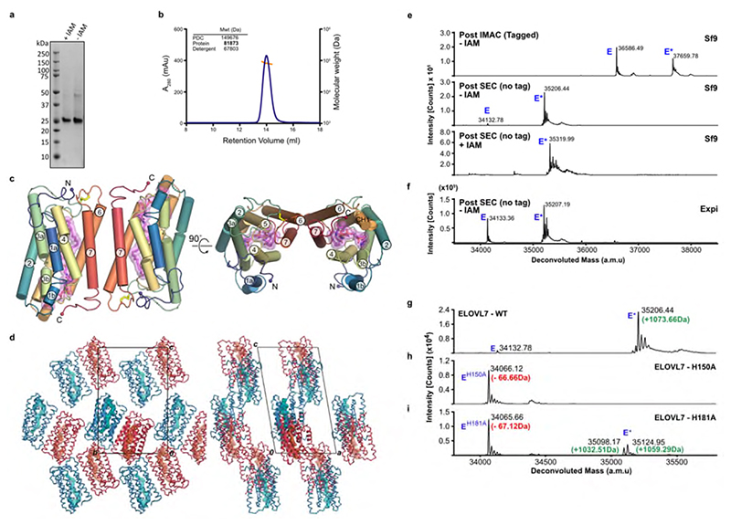
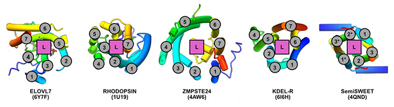
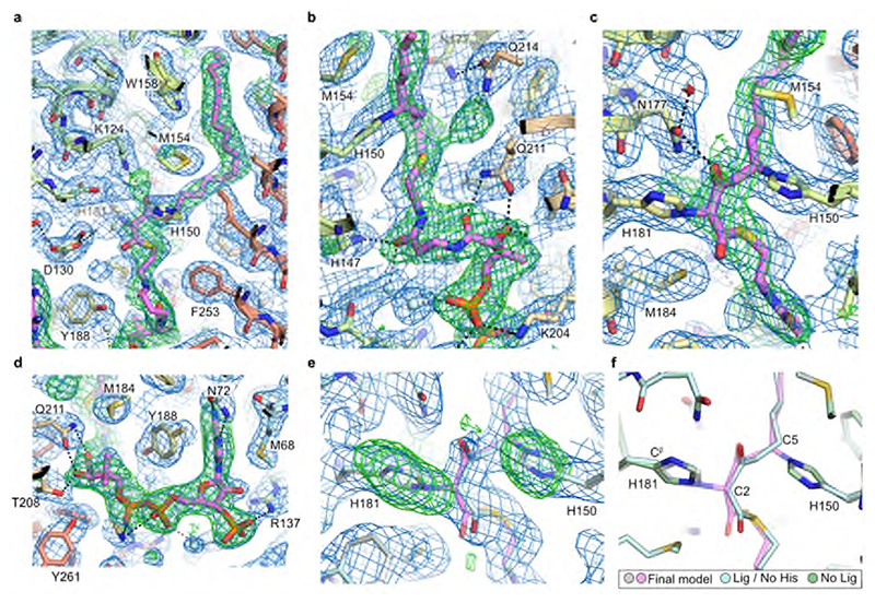
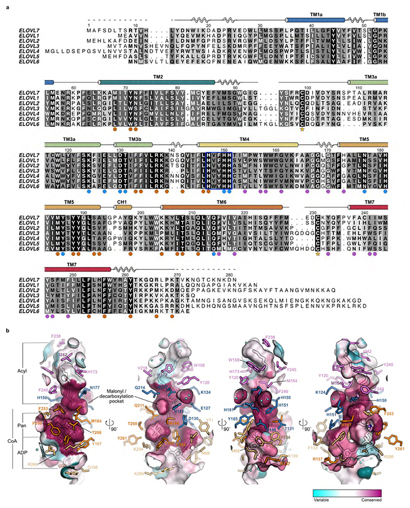
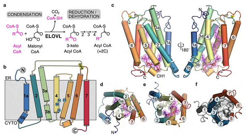
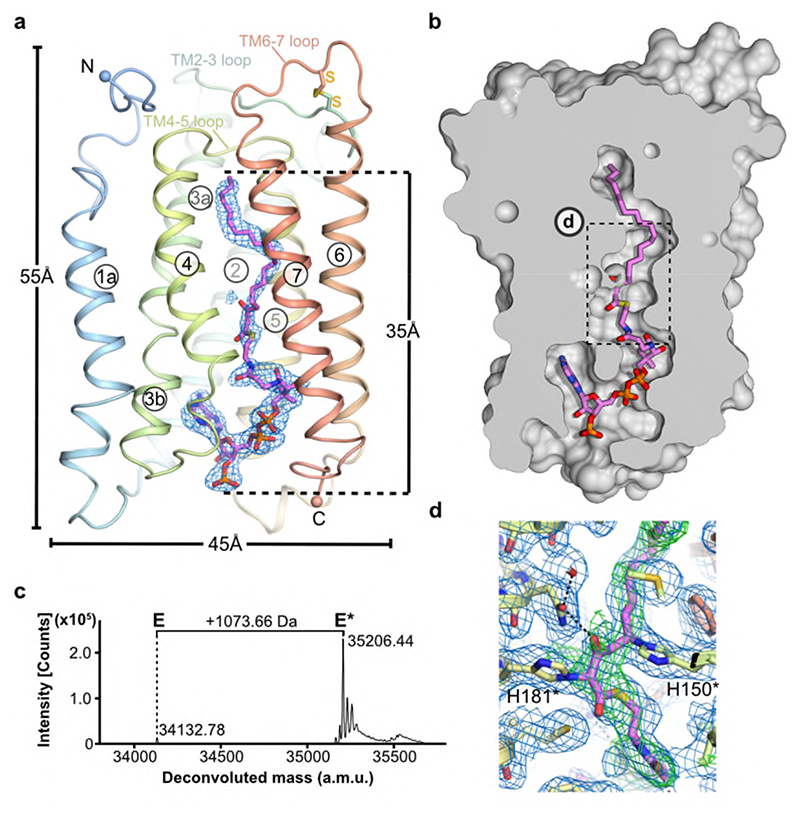
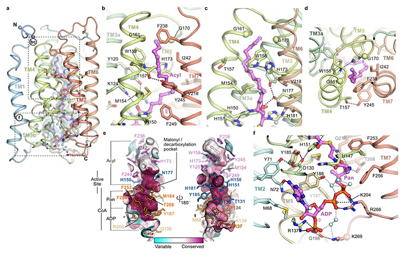
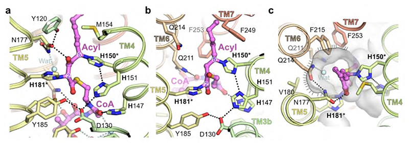
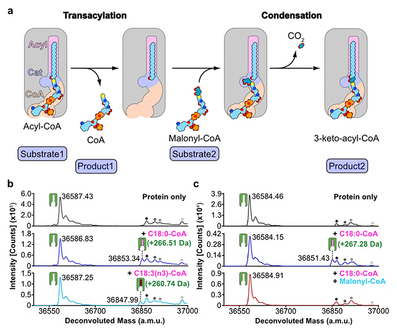
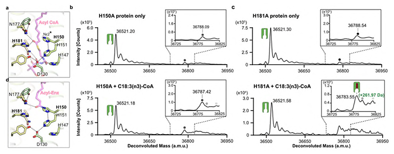
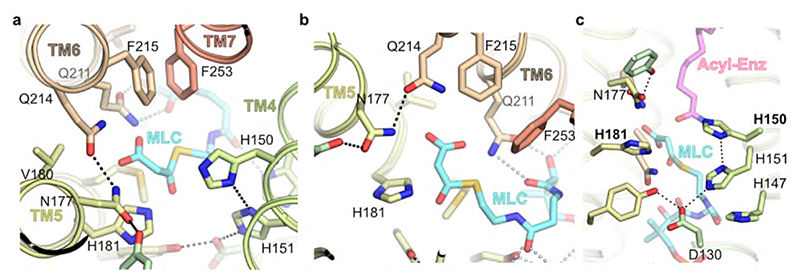
Very long chain fatty acids (VLCFAs) are essential building blocks for the synthesis of ceramides and sphingolipids. The first step in the fatty acid elongation cycle is catalyzed by the 3-keto acyl-coenzyme A (CoA) synthases (in mammals, ELOVL elongases). Although ELOVLs are implicated in common diseases, including insulin resistance, hepatic steatosis and Parkinson's, their underlying molecular mechanisms are unknown. Here we report the structure of the human ELOVL7 elongase, which comprises an inverted transmembrane barrel surrounding a 35-Å long tunnel containing a covalently attached product analogue. The structure reveals the substrate-binding sites in the narrow tunnel and an active site deep in the membrane. We demonstrate that chain elongation proceeds via an acyl-enzyme intermediate involving the second histidine in the canonical HxxHH motif. The unusual substrate-binding arrangement and chemistry suggest mechanisms for selective ELOVL inhibition, relevant for diseases where VLCFAs accumulate, such as X-linked adrenoleukodystrophy.


Conflict of interest statement

Competing Interests

The authors declare no competing interests.

Figures

Extended Data Fig. 1
Extended Data Fig. 1. Properties of purified ELOVL7.
a, SDS-PAGE gel of purified ELOVL7. Data shown is from a single purification but similar results were observed for multiple purifications / modifications. b, SEC profile and MALS analysis showing that OGNG-solubilised protein exists as a dimer in solution. Experiment carried out for tag-cleaved ELOVL7 purifications with and without IAM modification. Similar results were seen for each sample. c, Representation of head-to-tail dimer present in the crystal. d, ELOVL7 head-to-tail dimer packing within the crystal lattice. e, f, Intact mass analysis of ELOVL7 protein at various stages during purification. Deconvoluted mass spectra are shown for ELOVL7 protein purified from (e) Sf9 (n=3 or 4) and (f) Expi293F cells (n=2). For protein purified after expression in insect cells, the samples are shown after immobilized metal affinity chromatography (IMAC), after cleavage of the tag and size exclusion chromatography (SEC) and after treatment with iodoacetamide (IAM). The expected mass of the untagged ‘native’ enzyme (E) based on the sequence is 34222.38 Da. The observed mass peaks (Sf9, 34132.78 Da; Expi 34133.36 Da) correspond to the loss of the N-terminal methionine (-131.20 Da) and acetylation of the resulting new N-terminus (+42.04 Da). All samples were run in their reduced state. The modified material (E*) appears as an adduct with an average mass shift of +1073.6 Da. The addition of 113.55 Da upon treatment with IAM suggests modification of two cysteine residues. g-i, Deconvoluted intact mass spectra for the untagged g, WT (n=3), h, H150A (n=2) and i, H181A mutants (n=2). The expected mass decrease of a His-to-Ala mutation is 66.06 Da. Evidence of in vivo modification (E*) is observed for the H181A mutant but not for H150A. For intact mass experiments, theoretical and experimental masses along with mass errors are given in Supplementary Table 1.
Extended Data Fig. 2
Extended Data Fig. 2. TM helix topology of ELOVL7.
TM helical topology of ELOVL7 is compared with other six and seven membered TM bundles. TM helices are numbered and location of substrate/ligand site marked. Underlying cartoon representations of each structure are coloured from blue to red from the N- to C-termini respectively. PDB accession codes are shown in parentheses.
Extended Data Fig. 3
Extended Data Fig. 3. Electron density clearly shows covalently bound 3-keto-eicosanoyl-CoA.
a-d, Electron density running along the catalytic tunnel. Final BUSTER 2mFo-DFc (blue mesh, contoured at 1σ) and omit mFo-DFc (green mesh, contoured at 2.5σ) electron density maps are overlaid on the final model. e, Comparison of a test refinement in which the imidazole groups of H150 and H181 were removed from the model (grey carbon protein atoms / palecyan ligand carbon atoms) and the final model (palecyan protein carbons / violet ligand carbon atoms). The BUSTER 2mFo-DFc (blue mesh, contoured at 1σ) and mFo-DFc (green mesh, contoured at 3σ) maps for the refined histidine-truncated model / unlinked acyl-CoA are shown. f, Comparison of various refined models (green carbons - protein only; palecyan - protein without H150/181 sidechain plus ligand; grey/violet - final model with covalently attached ligand).
Extended Data Fig. 4
Extended Data Fig. 4. Sequence alignment and active site conservation of human ELOVL family members.
a, Sequence alignment of human ELOVL1-7. The conserved histidine box (147HxxHH151) is highlighted by a blue box. Filled circles below alignment indicate residues with a proposed catalytic role (blue) and residues interacting with either the CoA (orange) or acyl (plum) portion of the substrate. Cysteines that form the disulphide bridge (C99-C231) between the TM2/3 and TM6/7 loops are indicated by stars. b, Conservation of active site tunnel. Molecular surface representation is coloured by amino acid conservation score calculated by CONSURF analysis of a diverse set of ELOVL1-7 family members. The various subregions of the tunnel are indicated (ADP / Pan from CoA and Acyl chain). Amino acid residues that form the binding tunnel are coloured according to region (pink, acyl; blue, catalytic site; orange CoA binding).
Extended Data Fig. 5
Extended Data Fig. 5. WT ELOVL7 activity.
a,b Activity of residual WT enzyme on incubation with stearoyl-CoA (C18:0) and malonyl-CoA. Selected ion recording is shown for a, reaction mixture without added enzyme and b, reaction mixture after 3hr incubation with ELOVL7 enzyme. The ion peak at 1.61 mins corresponds to the expected 3-keto-eicosanoyl (C20)-CoA product of the elongation reaction. This experiment was carried out with two biological repeats with similar results.
Extended Data Fig. 6
Extended Data Fig. 6. Identification of a covalent acyl-enzyme intermediate of ELOVL7.
Purified, tagged, wildtype ELOVL7 was incubated in the presence and absence of known substrates and metalchelating agents prior to LC-ESI-MS intact mass analysis. a-g, Deconvoluted intact mass spectra for ELOVL7 incubated for 2h at 37°C. a, in the absence of substrates. b, ELOVL7 incubated with 100μM C18:0-CoA. Expected mass addition for acyl intermediate upon reaction with C18:0-CoA: +266.47 Da. c, ELOVL7 incubated with 100μM C18:3(n3)-CoA. Expected mass addition for acyl intermediate upon reaction with C18:3(n3)-CoA: +260.42 Da. d-e, ELOVL7 incubated with d, 100μM C18:0-CoA or e, 100μM C18:3(n3)-CoA in the presence of 1mM EDTA. f-g ELOVL7 incubated with f, 100μM C18:0-CoA or g, 100μM C18:3(n3)-CoA in the presence of 1mM EGTA. h-k, Sequential reaction of LMNG-purified ELOVL7 with C18:0-CoA and malonyl-CoA. h, LMNG-purified ELOVL in the absence of substrates. i, ELOVL7 incubated with 100μM C18:0-CoA. j, Purified ELOVL7 initially incubated with 100μM C18:0-CoA, followed by incubation with 200 μM malonyl-CoA. Addition of the second substrate leads to loss of the acyl-enzyme intermediate peak, consistent with the reaction having gone to completion. k, control ELOVL7 sample taken after incubation with C18:0-CoA was further incubated in the absence of malonyl-CoA, showing that covalent intermediate loss only occurs in the presence of malonyl-CoA. All experiments were repeated independently twice with similar results (n=2 biological repeats, see Supplementary Figure 1 and Figure 2 for replicate traces. See Supplementary Table 2 for theoretical and experimental masses and mass errors).
Extended Data Fig. 7
Extended Data Fig. 7. Proposed ping-pong reaction mechanism for ELOVL7.
a, Transacylation step with acyl chain of first substrate being transferred to H150. In the second step, malonyl-CoA binds and undergoes decarboxylation and a condensation reaction to form the elongated 3-keto product. b, Proposed reaction steps leading to C-N covalent adduct with H150 seen in crystal structure. In this scenario a 2,3-trans-enoyl-CoA serves as the first substrate (left) leading to the 3-keto,4,5-trans-enoyl-CoA ‘product’ (middle) which subsequently crosslinks to H150 via a conjugate addition reaction of H150 (right). The nature of the reaction that leads to H181 crosslinking to the C2 atom of the 3-keto-acyl-CoA is not clear.
Fig. 1
Fig. 1. Overall structure of ELOVL7.
a, FA elongase reaction sequence. b, Schematic representation of the seven transmembrane (TM) helix topology. c-e, Cartoon of ELOVL7 structure viewed c, parallel to the membrane plane and from either d, the ER or e, the cytoplasmic faces. f, ELOVL7 6-TM inverted barrel organisation viewed from the cytoplasmic face.
Fig. 2
Fig. 2. Heterologously expressed ELOVL7 is covalently bound to a 3-keto acyl-CoA.
a, Cartoon showing the covalently bound 3-keto eicosanoyl-CoA along with the FoFc omit electron density map (blue mesh, contoured at 3σ). b, Cutaway molecular surface representation showing the extent of the enclosed central active site tunnel. c, Intact mass analysis of Sf9 purified protein (E) highlighting adduct species (E*; +1073.66Da). Data shown from one experiment. Similar adduct peaks were observed for all purifications tested (n=3). d, Electron density in region around covalent linkages to active site histidines. Final BUSTER 2mFo-DFc (blue mesh, contoured at 1sigma) and omit mFo-DFc (green mesh, contoured at 2.5sigma) electron density maps are overlaid on the final model.
Fig. 3
Fig. 3. Acyl chain and CoA binding sites.
a, Overview of the central acyl-CoA binding tunnel. b-d, Acyl chain binding pocket. e, Conservation of active site tunnel. Molecular surface representation is coloured by amino acid conservation score calculated by CONSURF analysis of a diverse set of ELOV1-7 family members. The various subregions of the tunnel are indicated (ADP / Pan from CoA and Acyl chain). Amino acid residues that form the binding tunnel are coloured according to region (pink, acyl; blue, catalytic site; orange CoA binding). f, Cytoplasmic-facing CoA 3’-phospho ADP/pantothenic (Pan) binding pocket.
Fig. 4
Fig. 4. Catalytic site around the HxxHH motif.
a,b, Orthogonal views of central active site. c, Putative malonyl binding site adjacent to catalytic histidines. Molecular surface is shown in transparent grey highlighting the putative binding site for malonyl moiety of second substrate (indicated with hatched line). In the crystal structure this is filled by a water molecule (Wat). Covalently modified histidines (H150/H181) are highlighted by asterisks.
Fig. 5
Fig. 5. Proposed ping-pong mechanism and evidence for a covalent acyl-enzyme intermediate.
a, Schematic outlining proposed ELOVL ping-pong mechanism. b-c, Intact mass analysis of ELOVL7 in presence of b, acyl-CoA substrates demonstrating transacylation to form an acyl-enzyme intermediate and c, acyl-CoA followed by malonyl-CoA. Intact mass analysis of protein only (black), protein + C18:0 acyl-CoA (blue), protein + C18:3 acyl-CoA (cyan) and protein + C18:0 acyl-CoA + malonyl-CoA (red). Peaks are indicated as follows: protein (green icon), acyl enzyme intermediate (green icon + pink/red oblong), background species present in all traces (grey circles). (n=2 biological repeats, see Supplementary Figures 1-2 for replicate traces; Supplementary Table 2 for experimental and theoretical masses and mass errors).
Fig. 6
Fig. 6. Covalent acyl-enzyme intermediate is formed upon substrate reaction at H150.
a, Structural model for acyl-CoA binding. b-c, LC-ESI-MS intact mass analysis of mutant proteins upon incubation at 37°C for 2h. b, H150A mutant protein incubated in the absence of substrate (upper panel) or in the presence of 100 μM C18:3(n3)-CoA (lower panel). No acyl-enzyme intermediate formation could be detected with the H150A mutant protein. c, H181A mutant protein incubated in the absence of substrate (upper) or in the presence of 100μM C18:3(n3)-CoA (lower). Expected mass shift upon reaction with C18:3(n3)-CoA: +260.42 Da. The presence of a background peak at ~36788 Da precluded testing with C18:0-CoA. All intact mass experiments were repeated twice with similar results (n=2 biological repeats, see Supplementary Figure 3 for replicate traces; Supplementary Table 2 for experimental and theoretical masses and mass errors). Peaks are indicated as follows: protein (green icon), acyl enzyme intermediate (green icon + pink/red oblong), background species present in all traces (grey circles). d, Structural model for acyl-imidazole H150 intermediate. Ligand complexes are models based on the product analogue crystal structure. The unprotonated imidazole nitrogen of H150 involved in the transacylation step is highlighted.
Fig. 7
Fig. 7. Model for malonyl-CoA binding at the decarboxylation site.
a, Putative structural model for malonyl-CoA (MLC) binding within the conserved decarboxylation side pocket off the main tunnel. b, Conserved sidechains recognise malonyl carboxylate (viewed looking from H150). c, Composite structural model of second condensation reaction step with bound acy-limidazole intermediate and malonyl-CoA.

References

    1. Leonard AE, Pereira SL, Sprecher H, Huang Y-S. Elongation of long-chainfatty acids. Progress in Lipid Research. 2004;43:36–54. - PubMed
    1. Sassa T, Kihara A. Metabolism of very long-chain Fatty acids: genes and pathophysiology. Biomol Ther (Seoul) 2014;22:83–92. - PMC - PubMed
    1. Ohno Y, et al. ELOVL1 production of C24 acyl-CoAs is linked to C24 sphingolipid synthesis. Proc Natl Acad Sci U S A. 2010;107:18439–44. - PMC - PubMed
    1. Imgrund S, et al. Adult ceramide synthase 2 (CERS2)-deficient mice exhibit myelin sheath defects, cerebellar degeneration, and hepatocarcinomas. J Biol Chem. 2009;284:33549–60. - PMC - PubMed
    1. Isokawa M, Sassa T, Hattori S, Miyakawa T, Kihara A. Reduced chain length in myelin sphingolipids and poorer motor coordination in mice deficient in the fatty acid elongase Elovl1. FASEB Bioadv. 2019;1:747–759. - PMC - PubMed

Publication types

MeSH terms

LinkOut - more resources